 KSOEM
KSOEM

Articles
- Page Path
- HOME > Ann Occup Environ Med > Volume 38; 2026 > Article
- Case Report Basal cell carcinoma and squamous cell carcinoma of the hands in an orthopedic surgeon with occupational radiation exposure from C-arm fluoroscopy in Korea: a case report
-
Jisue Yoon1
 , Jihye Lee2, Seongjin Jeong1, Sangchul Roh1, Min-Gi Kim1, Jeongbae Rhie1,*
 -
Annals of Occupational and Environmental Medicine 2026;38:e25.
DOI: https://doi.org/10.35371/aoem.2026.38.e25
Published online: July 7, 2026
1Department of Occupational and Environmental Medicine, Dankook University Hospital, Cheonan, Korea
2Occupational Safety and Health Research Institute, Ulsan, Korea
- *Corresponding author: Jeongbae Rhie Department of Occupational and Environmental Medicine, Dankook University Hospital, 201 Manghyang-ro, Dongnam-gu, Cheonan 31116, Korea E-mail: rhie76@gmail.com
• Received: April 23, 2026 • Revised: June 27, 2026 • Accepted: June 28, 2026
© 2026 Korean Society of Occupational & Environmental Medicine
This is an Open Access article distributed under the terms of the Creative Commons Attribution Non-Commercial License (https://creativecommons.org/licenses/by-nc/4.0/) which permits unrestricted non-commercial use, distribution, and reproduction in any medium, provided the original work is properly cited.
- 815 Views
- 32 Download


Abstract
-
Background Occupational radiation exposure among medical professionals who use fluoroscopic devices is a well-recognized concern. While radiation-induced skin cancer has been reported in interventional radiologists, cases involving orthopedic surgeons with prolonged C-arm fluoroscopy exposure have rarely been described. We present a case of an orthopedic surgeon who developed basal cell carcinoma (BCC) and squamous cell carcinoma (SCC) on the hands after approximately 30 years of occupational radiation exposure from C-arm use during orthopedic surgeries in Korea.
-
Case presentation A 62-year-old male orthopedic surgeon who performed C-arm fluoroscopy–assisted surgeries for 30 years without radiation-protective gloves presented with bleeding and skin lesions on his left hand. Hospital records documented an average of 314 C-arm–assisted surgeries per year with an estimated annual C-arm usage of 12,397 minutes. An on-site radiation assessment was conducted in the actual operating room using a simulated patient, with positioning replicated via structured interview. Measurements using an X/gamma survey meter at the surgeon’s hand position yielded a mean equivalent dose rate of 3.5 mSv/h and a maximum of 5.0 mSv/h, corresponding to estimated annual equivalent doses of 723 and 1,033 mSv/y, both exceeding the regulatory equivalent dose limit for extremities in Korea. The patient had experienced recurrent blistering on both hands for approximately 10 years. Biopsy revealed BCC on the left thumb and SCC on the left second and third fingers, with bilateral chronic radiation dermatitis. Excision and skin grafting were performed on the affected fingers.
-
Conclusions This case demonstrates that prolonged C-arm fluoroscopy exposure without adequate hand protection can lead to radiation-induced skin cancers in orthopedic surgeons. The marked discrepancy between trunk-mounted dosimeter readings and estimated hand doses underscores the need for extremity-specific dosimetry. Radiation-protective gloves and regular dermatologic surveillance are essential for medical professionals exposed to ionizing radiation during fluoroscopy-guided procedures.
BACKGROUND
Skin cancer is broadly classified into malignant melanoma and non-melanoma skin cancer (NMSC), with NMSC further subdivided into basal cell carcinoma (BCC) and squamous cell carcinoma (SCC). BCC tends to be locally invasive with rare metastasis, whereas SCC commonly arises from precursor lesions such as actinic keratosis or Bowen’s disease. Ultraviolet (UV) radiation is the principal risk factor for both, and other established risk factors include organ transplantation, human immunodeficiency virus infection, human papillomavirus infection, and radiation therapy.1 According to the 2023 Korean cancer registry statistics, skin cancers including BCC and SCC accounted for 8,297 of 288,613 new cancer cases (2.9%), with a crude incidence rate of 16.2 per 100,000. Most patients were aged 70 years or older.2
Ionizing radiation causes DNA damage through both direct energy transfer to biological macromolecules and indirect generation of free radicals from water molecules, leading to chromosomal aberrations that may result in cell death or, if unrepaired, mutations that can give rise to cancer.3,4 The biological effects of ionizing radiation are classified as deterministic and stochastic effects, with carcinogenesis being a representative stochastic effect for which the linear no-threshold model is the current consensus, assuming that cancer risk increases proportionally with dose without a safe threshold.3,5 The International Agency for Research on Cancer (IARC) classifies X-radiation and gamma-radiation as Group 1 carcinogens, with sufficient evidence of carcinogenicity for skin (BCC) among other cancer sites including the salivary glands, esophagus, stomach, colon, lung, bone, female breast, bladder, brain, thyroid, kidney, and leukemia.6
In the medical field, numerous professionals are occupationally exposed to ionizing radiation, particularly during minimally invasive procedures that frequently utilize intraoperative fluoroscopy. Radiation exposure during interventional fluoroscopy can be classified into primary exposure when the operator is positioned within the imaging field, scattered radiation from the patient’s tissues, and leakage radiation from the X-ray tube. When the X-ray tube is positioned overhead, the operator’s head, neck, and hands may be exposed to unattenuated high-intensity radiation.7
In Korea, the mean annual personal radiation dose for medical radiation workers was 0.40 mSv/y in 2020;8 however, personal dosimeters may not always be worn, and the reported doses are likely underestimated.9 Beyond personal dosimetry, biological dosimetry using chromosomal aberration analysis in peripheral blood lymphocytes can also estimate the absorbed dose,10 and a Korean study using this method demonstrated that interventional radiologists exhibited significantly higher rates of dicentric chromosomes than the general population, suggesting that occupational radiation exposure among Korean interventional medical workers is not negligible.11 Cohort studies of medical radiation workers in the United States have demonstrated significant associations between occupational radiation exposure and the incidence of leukemia, breast cancer, and skin cancer,12 and a Korean study of diagnostic radiation workers also reported that overall cancer risk increased significantly with higher mean annual exposure doses.13
While radiation-induced skin cancer has been well-documented in interventional radiologists, cases involving orthopedic surgeons who routinely use C-arm fluoroscopy have been less frequently reported. We present a case in which both BCC and SCC developed concurrently on the hands of a single orthopedic surgeon after approximately 30 years of occupational radiation exposure from C-arm fluoroscopy, accompanied by bilateral chronic radiation dermatitis, in Korea.
CASE PRESENTATION
A 62-year-old male orthopedic surgeon presented to a local hospital in October 2021 with a chief complaint of bleeding from the left thumb. He reported a history of recurrent blistering on both hands for approximately 10 years prior to presentation. His past medical history was notable for hypertension, diagnosed in April 2018. He was a former smoker and reported consuming approximately two glasses of alcoholic drinks per day. There was no family history of skin cancer or other malignancies. He had no known history of excessive UV exposure, arsenic exposure, immunosuppressive therapy, or exposure to polycyclic aromatic hydrocarbons.
The patient obtained board certification in orthopedic surgery in February 1991, and had been working as the sole orthopedic surgeon at a local secondary hospital since then, for approximately 30 years at the time of diagnosis. His primary duties consisted of outpatient consultations and orthopedic surgeries, averaging approximately three surgeries per day.
Hospital records from 2008 to 2021 documented a total of 6,768 surgeries (approximately 480 per year), of which 4,425 (65.0%) involved C-arm fluoroscopy (approximately 314 C-arm–assisted surgeries per year). The types of C-arm–assisted surgeries included open reduction and internal fixation (OR/IF), closed reduction and internal fixation, fixation procedures, hardware removal, and foreign body removal. OR/IF procedures required the longest average C-arm usage time at 70 minutes per surgery. The total estimated annual C-arm fluoroscopy time was 12,397 minutes (Table 1).
Prior to 1991, the patient had no occupational history involving radiation exposure. Between 1991 and 2008, hospital records on the number of surgeries were unavailable; however, the patient reported that his surgical workload and C-arm usage patterns were consistent throughout his career.
All orthopedic surgeries were performed in a dedicated operating room on the seventh floor of the hospital. The specifications of the C-arm devices used are summarized in Supplementary Table 1. Records of models used prior to 2012 were unavailable. The KMC-950 (used 2012–2015; GEMSS Healthcare, Paju, Korea) operated at a tube voltage of 40–125 kV and a fluoroscopy tube current of 0.5–5 mA, with an inherent filtration of 0.8 mm beryllium. The SPINEL 3G (used from 2015 onward; GEMSS Healthcare) was equipped with an X-ray tube (model E7833X, Canon Electron Tubes & Devices Co., Ltd., Tochigi, Japan) operating at a tube voltage of 40–120 kV and a fluoroscopy tube current of 0.2–10 mA, with an inherent filtration of 0.7 mm Al at 70 kV. The on-site radiation measurements were performed using the SPINEL 3G, the model in use at the time of the epidemiological investigation.
During surgery, the patient routinely wore a lead apron to protect the trunk. However, the hands and upper extremities remained unshielded. Although radiation-protective gloves were available, the patient consistently chose not to wear them because they were heavy and significantly reduced manual dexterity and tactile sensation. Given the nature of orthopedic surgery, continuous fluoroscopic imaging was frequently employed, during which the surgeon’s hands were positioned within or near the primary radiation field. In orthopedic surgery, sterile field integrity requires that the surgeon’s hands remain within the operative field during fluoroscopic activation; consequently, the hands are likely exposed to radiation throughout the entirety of each C-arm operation cycle, supporting the assumption that the measured dose rates are applicable to the full C-arm usage time.
An on-site radiation exposure assessment was conducted in December 2023 in the actual operating room of the hospital where the patient had performed his surgeries. A simulated patient was positioned on the operating table, and the five most frequently performed surgery types were selected based on the patient’s surgical records. For each surgery type, the surgeon’s body position and hand placement were replicated in accordance with his own description of his operative positioning, obtained through a structured interview. Radiation measurements were performed using an Eberline FH40F4 X/gamma survey meter (Thermo Fisher Scientific, Waltham, MA, USA) placed at the position of the surgeon’s hands during C-arm fluoroscopy of three representative anatomical sites: knee, foot, and hip joint. The C-arm was configured in the standard orientation used for the measured procedures, with the X-ray tube positioned below the operating table and the image intensifier above, directing the primary beam in a caudocranial direction. For each surgical site, the survey meter recorded continuously throughout one complete C-arm operation cycle, and five repeated measurements were performed per site; the mean and maximum values were calculated from these measurements. The results demonstrated a mean equivalent dose rate of 3.5 mSv/h and a maximum equivalent dose rate of 5.0 mSv/h across all surgical sites (Table 2). Because the height of the surgeon’s hands relative to the C-arm source was similar across all surgery types, comparable dose rates were assumed for procedures not directly measured. Measurements from the anterior and posterior aspects of the surgical site showed minimal attenuation, indicating that patient tissue did not meaningfully reduce the radiation reaching the surgeon’s hands (Fig. 1). The survey meter had been calibrated by ENVIRO KOREA Co., Ltd. (Daejeon, Korea) on February 16, 2023, within 12 months prior to the measurements.
Based on the measured dose rates and estimated annual C-arm usage time, annual equivalent dose to the hands was calculated by multiplying the hourly dose rate by the estimated annual C-arm operating time (Table 3). Two exposure scenarios were considered: a base-case scenario using the mean dose rate of 3.5 mSv/h, yielding an estimated annual equivalent dose of 723 mSv/y, and an upper-bound scenario using the maximum dose rate of 5.0 mSv/h, yielding 1,033 mSv/y. Both values substantially exceeded the regulatory annual equivalent dose limit for extremities of radiation workers in Korea (500 mSv/y). It should be noted that these estimates are based on measurements conducted with C-arm equipment in use at the time of the investigation (2023), and that radiation output characteristics of earlier C-arm models used during the patient’s career may have differed.
The patient’s personal dosimeter records from 2017 to 2022 were reviewed (Supplementary Table 2). The trunk-mounted dosimeter recorded annual cumulative doses of 0.03–0.34 mSv. Additional measurements at the dosimeter position during C-arm operation showed approximately 0.16 mSv/h. This marked discrepancy between trunk and hand doses reflects the differential shielding provided by the lead apron and direct exposure of unprotected hands to the radiation field (Supplementary Fig. 1).
Physical examination revealed bilateral skin changes consistent with chronic radiation dermatitis, including diffuse erythema, dryness, scaling, skin atrophy, telangiectasia, and nail dystrophy (Fig. 2). On the left thumb, a skin-colored raised lesion with erythematous patches and spontaneous bleeding from telangiectatic vessels was observed (Fig. 3). On the left second and third fingers, erythematous patches with characteristic scaly crusts were noted (Fig. 4). On the right second finger, a protruding lesion accompanied by telangiectasia was also observed (Supplementary Fig. 2).
Based on these findings, the patient was diagnosed with bilateral chronic radiation dermatitis of the hands. Because the lesions on the left hand were highly suspicious for skin malignancy, the patient was referred to a tertiary hospital, where punch biopsy was performed on November 11, 2021. Histopathological examination revealed BCC on the left thumb and well-differentiated SCC on the left second and third fingers. Preoperative laboratory tests, chest X-ray, and chest computed tomography showed no other abnormal findings. Magnetic resonance imaging of the left hand demonstrated lesions consistent with the biopsy-confirmed malignancies.
In December 2021, the patient underwent surgical treatment, consisting of wide excision with full-thickness skin grafting for the left thumb (BCC) and wide tumor excision for the left third finger (SCC). The remaining lesions were treated with topical 5-fluorouracil (Efudex, Bausch Health Canada Inc., Laval, QC, Canada) cream and cryotherapy. Intraoperative histopathological examination confirmed the same diagnoses as the punch biopsy, and all lesions were completely excised with negative resection margins and without evidence of lymphovascular or perineural invasion. The patient continued to be followed for monitoring of the remaining lesions.
This study was approved by the Institutional Review Board of Dankook University (IRB No. DKU 2025-12-021-002). Informed consent was obtained from the patient for the publication of this case report and accompanying images.
DISCUSSION AND CONCLUSION
We report a case of BCC and SCC of the hands in an orthopedic surgeon who was occupationally exposed to ionizing radiation from C-arm fluoroscopy for approximately 30 years without adequate hand protection. The estimated annual equivalent dose to his hands (723–1,033 mSv/y) far exceeded the regulatory dose limits, and the development of multiple skin cancers accompanied by bilateral chronic radiation dermatitis strongly supports a causal relationship with occupational radiation exposure.
The causal relationship between occupational radiation exposure and skin cancer in this patient is supported by several lines of evidence: (1) Intensity and duration of exposure: the estimated annual equivalent dose (723–1,033 mSv/y) over approximately 30 years far exceeded regulatory limits; (2) Latency: skin cancers developed after a latency period of approximately 30 years, consistent with radiation-induced solid tumors; (3) Anatomical concordance: malignancies arose exclusively on the hands, the body region with the highest occupational radiation exposure; (4) Precursor lesion: bilateral chronic radiation dermatitis preceded the malignancies, a well-established precursor to radiation-induced skin cancer; and (5) Exclusion of alternative risk factors: no significant UV exposure, arsenic exposure, immunosuppressive therapy, or relevant family history was identified.
The IARC classifies X-radiation and gamma-radiation as Group 1 carcinogens, with sufficient evidence for BCC of the skin among other cancers.6 Studies of Japanese atomic bomb survivors demonstrated a dose-response relationship between radiation exposure and BCC, although such a relationship was not confirmed for SCC.14 A 2007 follow-up study of the same cohort also failed to demonstrate an association for SCC.15 In a study of 10,834 individuals irradiated for tinea capitis in childhood, the predominant skin cancer was BCC, with zero cases of SCC identified in the exposed group compared to two in the control group. The investigators noted that BCC may arise from mild to moderate radiation exposure, whereas SCC tends to occur after high-intensity local radiation exposure, often accompanied by radiation dermatitis, and that BCC predominantly affects the head and neck region while SCC more commonly develops on the distal extremities.16 A 50-year follow-up study of tinea capitis patients similarly found that the few SCCs in the irradiated group mostly co-occurred with BCC.17
A U.S. study of 65,304 radiologic technologists found a significant trend of increasing BCC risk with earlier year of first employment, but no such trend for SCC.18 A U.S. case-control study by Lichter et al.19 found that therapeutic ionizing radiation increased the risk of both BCC and SCC at irradiated sites, and suggested that fractionated moderate-dose radiation may be more carcinogenic than single high-dose exposures.20
Although the epidemiological evidence for radiation-induced SCC is less robust than for BCC, and the mechanisms by which radiation exposure differentially affects BCC and SCC pathogenesis remain unclear,21 historically, the first cancer attributed to radiation was an SCC on the hand of a radiation worker reported in 1902.22 Numerous early radiation workers who were chronically exposed to high-intensity local radiation developed SCC accompanied by radiation dermatitis and ulceration on the upper extremities.23 In the present case, SCC developed on fingers with chronic radiation dermatitis following prolonged high-dose local radiation exposure, which is consistent with these historical observations.
Chronic radiation dermatitis is a deterministic effect of ionizing radiation, occurring when cumulative doses exceed a threshold sufficient to cause progressive tissue damage. Unlike stochastic effects such as carcinogenesis, deterministic effects have a dose threshold and increase in severity with dose. The bilateral chronic radiation dermatitis observed in this patient, with features of skin atrophy, telangiectasia, and nail dystrophy, serves as a biological indicator of sustained high-dose radiation exposure to the hands and provides independent corroboration of the estimated doses exceeding regulatory limits.
Physicians performing interventional procedures are continuously exposed to high-intensity radiation on their hands.24 A Korean investigation documented SCC on the second finger of a 52-year-old interventional radiologist after more than 37,000 procedures over 11 years, preceded by chronic radiation dermatitis.25 Jang et al.26 and Kim et al.27 similarly reported finger SCC in radiologists with long-term procedural exposure, each preceded by chronic radiation dermatitis.
Among orthopedic surgeons, Yoon et al.28 reported SCC on the fingers of an orthopedic surgeon who performed approximately 700 percutaneous vertebral augmentation procedures and approximately 70,000 nerve blocks over 11 years. The present case is notable in that the primary source of radiation exposure was routine C-arm fluoroscopy during general orthopedic procedures (fracture fixation, hardware removal, etc.) rather than specific interventional procedures, suggesting that routine orthopedic surgery involving C-arm use can also pose a substantial cumulative radiation risk to the hands.
It should be noted that this patient’s workload of approximately 314 C-arm–assisted surgeries per year is substantial but not exceptional for a sole orthopedic surgeon at a secondary hospital in Korea. While this case may represent a high-exposure scenario, it is likely that many orthopedic surgeons with similar surgical volumes and prolonged careers may face comparable cumulative radiation exposure to the hands, particularly if they do not consistently use radiation-protective gloves. This case therefore has broader implications for occupational health screening and radiation protection practices among orthopedic surgeons who routinely use C-arm fluoroscopy.
A particularly significant finding is the marked discrepancy between the personal dosimeter readings and the estimated annual equivalent doses. This illustrates the inadequacy of trunk-mounted dosimeters in reflecting extremity doses for surgeons whose hands are routinely positioned within the C-arm radiation field, consistent with reports that personal dosimeter readings are likely underestimated.9 Regulatory bodies should therefore consider mandating ring dosimetry for hand dose monitoring for all physicians who perform fluoroscopy-guided procedures.
Beyond dosimetry, the present case also reveals a critical gap in the occupational health surveillance system. The patient had undergone periodic special health examinations as a radiation worker, and records confirmed that annual examinations conducted from 2016 through March 2021 consistently documented no significant dermatologic findings. Yet during this same period, the patient had been experiencing recurrent blistering on both hands—a symptom he later reported had persisted for approximately 10 years prior to diagnosis. This discrepancy highlights a limitation of the current special health examination framework for radiation workers in Korea. The reason these lesions were not detected earlier cannot be determined with certainty from the available records; the patient may not have reported the intermittent blistering during examinations, or routine examinations may not have included detailed inspection of the hands. Regardless of the specific cause, this case illustrates that current surveillance practices may be insufficient to detect early cutaneous changes in radiation workers. We recommend that the dermatologic component of special health examinations for radiation workers who routinely expose the hands to radiation be revised to include systematic visual inspection of the hands and forearms by a physician, with established referral criteria for dermatologic consultation. Such a practical revision could enable earlier detection of radiation-induced skin changes and prevent progression to malignancy. More broadly, this case reflects a systemic failure extending beyond health surveillance alone: hand exposure was never quantified owing to the absence of extremity dosimetry, direct exposure continued without enforced hand protection, and the surveillance system failed to detect the resulting skin changes. Together, these underscore the need for an integrated approach encompassing exposure monitoring, protective equipment enforcement, and targeted health surveillance.
Regarding other potential risk factors for skin cancer, UV radiation is the most well-established cause of cutaneous SCC,29 and occupational UV exposure approximately doubles the risk.30 However, the patient worked primarily indoors in an operating room with no significant occupational or recreational UV exposure. Other risk factors including polycyclic aromatic hydrocarbons, arsenic, and immunosuppressive agents were not identified in this patient’s history.
Although the patient was 62 years old at diagnosis and aging itself increases skin cancer risk, the bilateral distribution limited to the hands—the body region with the highest occupational radiation exposure—argues against age-related skin cancer, which typically affects sun-exposed areas such as the face and scalp. The patient was a former smoker; however, smoking has not been established as an independent risk factor for non-melanoma skin cancer. Regarding incidental UV exposure to the hands during daily activities, the patient’s work was predominantly indoors, and he reported no significant outdoor recreational activities. The anatomical specificity of the lesions to the hands, combined with the coexistence of radiation dermatitis bilaterally, strongly points to occupational radiation as the predominant etiological factor.
This case has a limitation in that the exact cumulative radiation dose over the patient’s entire career could not be precisely determined, because C-arm records prior to 2012 and surgical volume data prior to 2008 were unavailable. Several additional uncertainties arise from reconstructing exposure over a 30-year period using contemporary measurements. The two scenarios presented reflect measurement variability rather than historical variability in C-arm technology and fluoroscopy practice. Notably, the output characteristics of older C-arm systems could not be verified, and historical exposures may therefore have differed from the present measurements in either direction. Furthermore, the assumption that the hands remained within the radiation field throughout the entire fluoroscopy time, although supported by the requirements of sterile surgical technique, simplifies actual practice. Nevertheless, the radiation exposure estimated from the period for which records were available substantially exceeded the regulatory dose limit for extremities, which is sufficient to support a causal relationship between occupational radiation exposure and the development of skin cancer in this patient.
In conclusion, this case, consistent with previous reports, demonstrates that chronic high-intensity occupational radiation exposure is associated with the development of not only BCC but also SCC. Even when personal dosimeters record low cumulative exposure, radiation doses to body regions not adequately shielded by lead aprons, such as the hands, may be substantially underestimated. Therefore, the consistent use of radiation-protective gloves is essential for medical professionals who use C-arm fluoroscopy to prevent radiation-induced skin cancer, and regular dermatologic surveillance for the early detection of precancerous lesions is also warranted. In addition, hospitals should ensure an adequate supply of radiation protective equipment, including lead aprons, thyroid shields, and radiation-protective gloves, and should implement in-hospital educational programs to train medical workers on the proper use of protective equipment and to emphasize the risks of occupational radiation exposure.
Abbreviations
BCC
basal cell carcinoma
CR/IF
closed reduction and internal fixation
FB
foreign body
IARC
International Agency for Research on Cancer
NMSC
non-melanoma skin cancer
OR/IF
open reduction and internal fixation
SCC
squamous cell carcinoma
UV
ultraviolet
-
Competing interests
No potential conflict of interest relevant to this article was reported.
-
Author contributions
Conceptualization: Yoon J, Rhie J. Data curation: Yoon J, Lee J, Jeong S, Kim MG. Methodology/formal analysis/validation: Yoon J, Lee J, Jeong S, Roh S, Rhie J. Project administration: Rhie J. Writing - original draft: Yoon J. Writing - review & editing: Yoon J, Lee J, Jeong S, Roh S, Kim MG, Rhie J.
-
Acknowledgments
This study was conducted as a commissioned epidemiological investigation by the Occupational Safety and Health Research Institute (OSHRI) pursuant to Article 43-2 of the Occupational Safety and Health Act of Korea (investigation period: March 14, 2023–March 15, 2024).
NOTES
SUPPLEMENTARY MATERIAL
Supplementary Table 1.
Specifications of C-arm devices used in the operating room.
Supplementary Table 2.
Annual cumulative radiation doses from the personal dosimeter (trunk-mounted).
Supplementary Fig. 1.
Radiation dose rate at the trunk-mounted personal dosimeter position, showing approximately 0.16 mSv/h.
Supplementary Fig. 2.
Right hand showing chronic radiation dermatitis lesion.
Fig. 1.
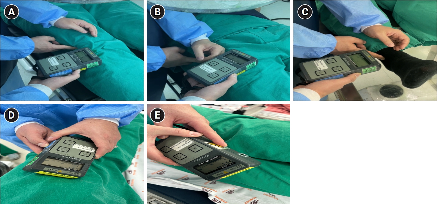
On-site radiation dose measurement at the surgeon's hand position during C-arm fluoroscopy. (A) Knee surgery. (B) Hip joint surgery. (C) Foot surgery. (D, E) Anterior and posterior aspects of the surgical site, demonstrating minimal attenuation through the patient's tissues. Measurements were obtained at 30–40 cm from the X-ray source under simulated surgical conditions.
Fig. 2.
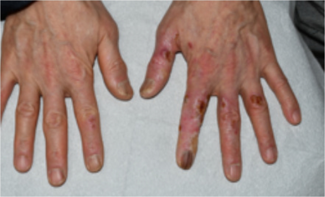
Both hands of the patient showing bilateral chronic radiation dermatitis. Presenting diffuse erythema, dryness, scaling, skin atrophy, telangiectasia, and nail dystrophy. Photographs were taken during the epidemiological investigation in December 2023.
Fig. 3.
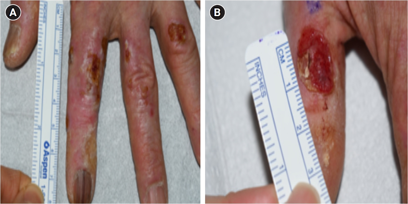
Close-up view of the left hand. (A) Overview of the left hand showing chronic radiation dermatitis. (B) Close-up of the left thumb showing a skin-colored raised lesion with erythematous patches and spontaneous bleeding from telangiectatic vessels, subsequently diagnosed as basal cell carcinoma on punch biopsy.
Fig. 4.
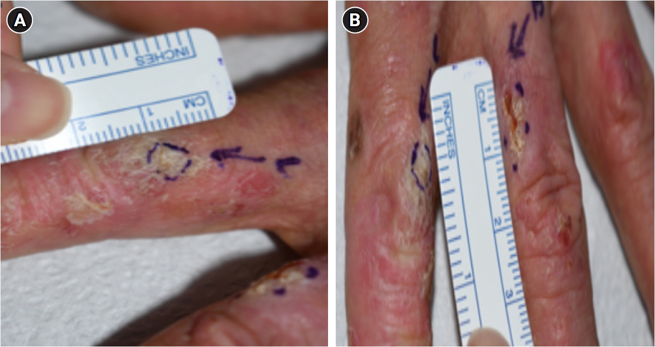
Close-up view of the left second and third fingers. Second finger (A) and third finger (B) showing erythematous patches with characteristic scaly crusts, subsequently diagnosed as well-differentiated squamous cell carcinoma on punch biopsy.
Table 1.
Types of C-arm–assisted surgeries and C-arm usage time
Table 2.
Radiation dose rates measured at the surgeon’s hand position during C-arm use
| Surgical site | Maximum (mSv/h) | Average (mSv/h) |
|---|---|---|
| Knee | 5.0 | 3.5 |
| Foot | 5.5 | 4.0 |
| Hip joint | 4.5 | 3.0 |
| Average | 5.0 | 3.5 |
Table 3.
Estimated annual equivalent dose
| Method | Formula | Annual equivalent dose (mSv/y) |
|---|---|---|
| Average dose rate | 3.5 mSv/h × 12,397 min/y × 1 h/60 min | 723 |
| Maximum dose rate | 5.0 mSv/h × 12,397 min/y × 1 h/60 min | 1,033 |
- 1. Gordon R. Skin cancer: an overview of epidemiology and risk factors. Semin Oncol Nurs 2013;29(3):160–9.ArticlePubMed
- 2. Korea Central Cancer Registry. Cancer incidence by 61 cancer types, sex, and age group (5-year). https://kosis.kr. Updated 2026. Accessed March 31, 2026.
- 3. Shah DJ, Sachs RK, Wilson DJ. Radiation-induced cancer: a modern view. Br J Radiol 2012;85(1020):e1166–73.ArticlePubMedPMC
- 4. National Research Council. Health Risks from Exposure to Low Levels of Ionizing Radiation: BEIR VII Phase 2. Washington, DC: National Academies Press; 2006.
- 5. Boice JD Jr. The linear nonthreshold (LNT) model as used in radiation protection: an NCRP update. Int J Radiat Biol 2017;93(10):1079–92.ArticlePubMed
- 6. International Agency for Research on Cancer. List of classifications by cancer sites with sufficient or limited evidence in humans, IARC Monographs Volumes 1–140. https://monographs.iarc.who.int/wp-content/uploads/2019/07/Classifications_by_cancer_site.pdf. Updated 2025. Accessed March 31, 2026.
- 7. Schueler BA. Operator shielding: how and why. Tech Vasc Interv Radiol 2010;13(3):167–71.ArticlePubMed
- 8. Korea Disease Control and Prevention Agency. 2020 Annual Report on Personal Radiation Doses of Radiation Workers in Medical Institutions. Cheongju, Korea: Korea Disease Control and Prevention Agency; 2021.
- 9. Lee WJ. Occupational radiation exposure and health effects among medical workers. Korean J Med 2018;93(3):237–46.ArticlePDF
- 10. Jeong TS, Baik HM, Shin BC, Moon CW, Kim MH, Lee YH, et al. The frequency of chromosomal aberrations in peripheral lymphocytes according to radiation dose and dose rate. J Korean Soc Ther Radiol Oncol 2000;18(2):138–49.
- 11. Lee Y, Lee WJ, Jin YW, Jang S. Interventional radiologists have a higher rate of chromosomal damage due to occupational radiation exposure: a dicentric chromosome assay. Eur Radiol 2021;31(11):8256–63.ArticlePubMedPDF
- 12. Yoshinaga S, Mabuchi K, Sigurdson AJ, Doody MM, Ron E. Cancer risks among radiologists and radiologic technologists: review of epidemiologic studies. Radiology 2004;233(2):313–21.ArticlePubMed
- 13. Choi KH, Ha M, Lee WJ, Hwang SS, Jeong M, Jin YW, et al. Cancer risk in diagnostic radiation workers in Korea from 1996–2002. Int J Environ Res Public Health 2013;10(1):314–27.ArticlePubMedPMC
- 14. Ron E, Preston DL, Kishikawa M, Kobuke T, Iseki M, Tokuoka S, et al. Skin tumor risk among atomic-bomb survivors in Japan. Cancer Causes Control 1998;9(4):393–401.ArticlePubMedPDF
- 15. Preston DL, Ron E, Tokuoka S, Funamoto S, Nishi N, Soda M, et al. Solid cancer incidence in atomic bomb survivors: 1958–1998. Radiat Res 2007;168(1):1–64.ArticlePubMed
- 16. Ron E, Modan B, Preston D, Alfandary E, Stovall M, Boice JD Jr. Radiation-induced skin carcinomas of the head and neck. Radiat Res 1991;125(3):318–25.ArticlePubMed
- 17. Shore RE, Moseson M, Xue X, Tse Y, Harley N, Pasternack BS. Skin cancer after X-ray treatment for scalp ringworm. Radiat Res 2002;157(4):410–8.ArticlePubMed
- 18. Yoshinaga S, Hauptmann M, Sigurdson AJ, Doody MM, Freedman DM, Alexander BH, et al. Nonmelanoma skin cancer in relation to ionizing radiation exposure among U.S. radiologic technologists. Int J Cancer 2005;115(5):828–34.ArticlePubMed
- 19. Lichter MD, Karagas MR, Mott LA, Spencer SK, Stukel TA, Greenberg ER. Therapeutic ionizing radiation and the incidence of basal cell carcinoma and squamous cell carcinoma. The New Hampshire Skin Cancer Study Group. Arch Dermatol 2000;136(8):1007–11.ArticlePubMed
- 20. Goldschmidt H, Breneman JC, Breneman DL. Ionizing radiation therapy in dermatology. J Am Acad Dermatol 1994;30(2 Pt 1):157–82.ArticlePubMed
- 21. Li C, Athar M. Ionizing radiation exposure and basal cell carcinoma pathogenesis. Radiat Res 2016;185(3):217–28.ArticlePubMedPMC
- 22. Frieben A. Demonstration eines cancroids des rechten handruckens, das sich nach langedauernder einwirkung von rontgenstrahlen entwickelt hatte [Demonstration of a carcinoma (cancroid) of the dorsum of the right hand that developed after prolonged exposure to X-rays]. Fortschr Geb Rontgestr 1902;6:106–11.
- 23. Shore RE. Radiation-induced skin cancer in humans. Med Pediatr Oncol 2001;36(5):549–54.ArticlePubMed
- 24. Stavas JM, Smith TP, DeLong DM, Miller MJ, Suhocki PV, Newman GE. Radiation hand exposure during restoration of flow to the thrombosed dialysis access graft. J Vasc Interv Radiol 2006;17(10):1611–7.ArticlePubMed
- 25. Korea Occupational Safety and Health Agency. Casebook of Occupational Disease Diagnosis 2018–2019: Epidemiological Investigation of Skin Cancer in an Interventional Radiologist. Seoul, Korea: KOSHA; 2019, 37–8.
- 26. Jang S, Jin YW, Lee WJ. Estimating occupational radiation exposure using cytogenetic dosimetry in an interventional radiologist with skin cancer: a case report. J Vasc Interv Radiol 2020;31(11):1960–3.ArticlePubMed
- 27. Kim DH, Kim HS, Lee JY, Park HJ, Cho BK. A case of chronic radiodermatitis with CML and squamous cell carcinoma in radiologist. Ann Dermatol 2006;18(1):9–13.Article
- 28. Yoon HS, Heo JH, Lee SH, Shin J, Choi GS, Byun JW. Squamous cell carcinoma on the fingers of orthopedic surgeon induced by occupational radiation exposure. Ann Dermatol 2019;31(2):251–3.ArticlePubMedPMCPDF
- 29. Rigel DS. Cutaneous ultraviolet exposure and its relationship to the development of skin cancer. J Am Acad Dermatol 2008;58(5 Suppl 2):S129–32.ArticlePubMed
- 30. Schmitt J, Seidler A, Diepgen TL, Bauer A. Occupational ultraviolet light exposure increases the risk for the development of cutaneous squamous cell carcinoma: a systematic review and meta-analysis. Br J Dermatol 2011;164(2):291–307.ArticlePubMed
REFERENCES
Figure & Data
REFERENCES
Citations
Citations to this article as recorded by 
 Cite
Cite XML Download
Basal cell carcinoma and squamous cell carcinoma of the hands in an orthopedic surgeon with occupational radiation exposure from C-arm fluoroscopy in Korea: a case report
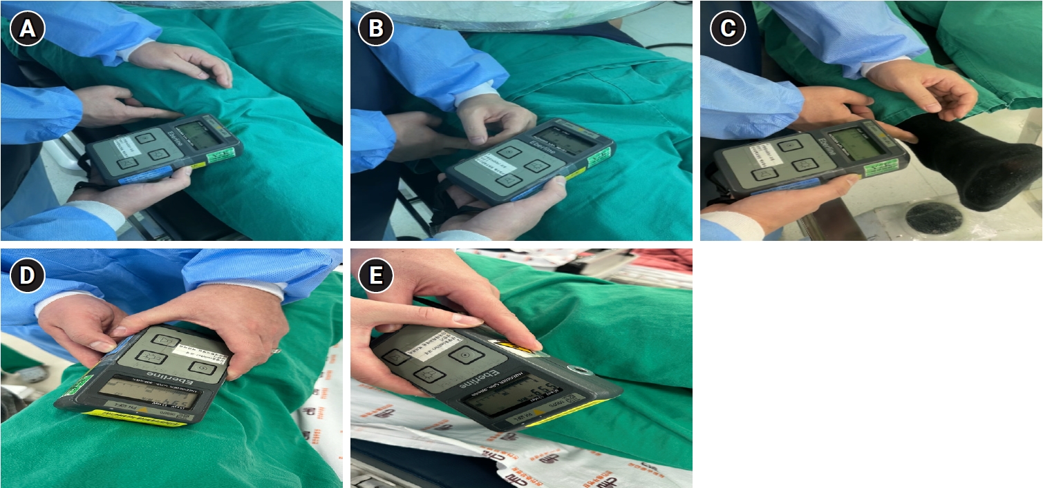
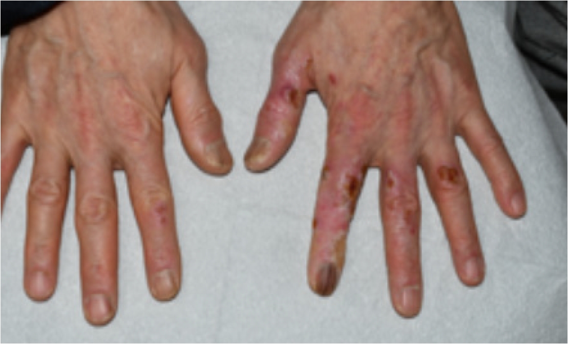
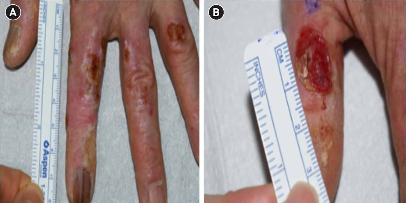
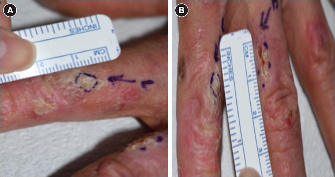
Fig. 1. On-site radiation dose measurement at the surgeon's hand position during C-arm fluoroscopy. (A) Knee surgery. (B) Hip joint surgery. (C) Foot surgery. (D, E) Anterior and posterior aspects of the surgical site, demonstrating minimal attenuation through the patient's tissues. Measurements were obtained at 30–40 cm from the X-ray source under simulated surgical conditions.
Fig. 2. Both hands of the patient showing bilateral chronic radiation dermatitis. Presenting diffuse erythema, dryness, scaling, skin atrophy, telangiectasia, and nail dystrophy. Photographs were taken during the epidemiological investigation in December 2023.
Fig. 3. Close-up view of the left hand. (A) Overview of the left hand showing chronic radiation dermatitis. (B) Close-up of the left thumb showing a skin-colored raised lesion with erythematous patches and spontaneous bleeding from telangiectatic vessels, subsequently diagnosed as basal cell carcinoma on punch biopsy.
Fig. 4. Close-up view of the left second and third fingers. Second finger (A) and third finger (B) showing erythematous patches with characteristic scaly crusts, subsequently diagnosed as well-differentiated squamous cell carcinoma on punch biopsy.
Fig. 1.
Fig. 2.
Fig. 3.
Fig. 4.
Basal cell carcinoma and squamous cell carcinoma of the hands in an orthopedic surgeon with occupational radiation exposure from C-arm fluoroscopy in Korea: a case report
| Type of surgery | C-arm time per surgery (min) | Annual surgeries | Annual C-arm time (min) | Proportion of annual C-arm time (%) |
|---|---|---|---|---|
| OR/IF | 70 | 135 | 9,450 | 76.2 |
| CR/IF | 13 | 33 | 429 | 3.5 |
| Fixation (nailing, pinning, wire) | 13 | 12 | 156 | 1.3 |
| Removal (pin, nail, wire, cable) | 18 | 124 | 2,232 | 18.0 |
| FB removal | 13 | 10 | 130 | 1.0 |
| Total | – | 314 | 12,397 | 100.0 |
| Surgical site | Maximum (mSv/h) | Average (mSv/h) |
|---|---|---|
| Knee | 5.0 | 3.5 |
| Foot | 5.5 | 4.0 |
| Hip joint | 4.5 | 3.0 |
| Average | 5.0 | 3.5 |
| Method | Formula | Annual equivalent dose (mSv/y) |
|---|---|---|
| Average dose rate | 3.5 mSv/h × 12,397 min/y × 1 h/60 min | 723 |
| Maximum dose rate | 5.0 mSv/h × 12,397 min/y × 1 h/60 min | 1,033 |
Table 1. Types of C-arm–assisted surgeries and C-arm usage time
OR/IF: open reduction and internal fixation; CR/IF: closed reduction and internal fixation; FB: foreign body.
Table 2. Radiation dose rates measured at the surgeon’s hand position during C-arm use
Table 3. Estimated annual equivalent dose


