
Articles
- Page Path
- HOME > Ann Occup Environ Med > Volume 35; 2023 > Article
- Original Article Environment-wide association study of elevated liver enzymes: results from the Korean National Environmental Health Survey 2018–2022
-
Youngchan Chi1
 , Jong-Tae Park1, Sewhan Na1,2, Kyeongmin Kwak1
 -
Annals of Occupational and Environmental Medicine 2023;35:e27.
DOI: https://doi.org/10.35371/aoem.2023.35.e27
Published online: July 31, 2023
1Department of Occupational and Environmental Medicine, Korea University Ansan Hospital, Ansan, Korea.
2Department of Environmental Health Sciences, Seoul National University Graduate School of Public Health, Seoul, Korea.
- Correspondence: Kyeongmin Kwak. Department of Occupational and Environmental Medicine, Korea University Ansan Hospital, 123 Jeokgeum-ro, Danwon-gu, Ansan 15355, Korea. pathfinder81@korea.ac.kr
• Received: December 21, 2022 • Revised: April 27, 2023 • Accepted: June 11, 2023
Copyright © 2023 Korean Society of Occupational & Environmental Medicine
This is an Open Access article distributed under the terms of the Creative Commons Attribution Non-Commercial License (https://creativecommons.org/licenses/by-nc/4.0/) which permits unrestricted non-commercial use, distribution, and reproduction in any medium, provided the original work is properly cited.
Abstract
-
Background Environmental exposure is characterized by low concentration, chronic, and complex exposure. Traditional epidemiological studies show limitations in reflecting these characteristics since they usually focus on a single or very limited number of exposure factors at a time. In this study, we adopted the methodology of environment-wide association study (EWAS) to figure out the association of human liver function with various environmentally hazardous substances.
-
Methods We analyzed 2,961 participants from the Korean National Environmental Health Survey Cycle 4 (2018–2020). Using generalized linear model (GLM) analysis, we analyzed the association of 72 variables with 3 liver function indices (aspartate aminotransferase [AST], alanine aminotransferase [ALT], and gamma glutamyl transferase [GGT]). Finally, we visualized our results with Manhattan plot.
-
Results In GLM analysis, perfluorooctanesulfonate were positively associated with ALT (odds ratio [OR]: 2.2; 95% confidence interval [CI]: 1.39–3.46; p adjusted = 0.0147) and perfluorodecanoic acid showed positive association with GGT (OR: 2.73; 95% CI: 1.36–5.5; p adjusted = 0.0256). Plasma mercury showed positive association with GGT (OR: 1.45; 95% CI: 1.14–1.84; p adjusted = 0.0315). Using a plastic container while keeping food in the refrigerator was associated with elevated GGT compared to using a glass container (OR: 1.51; 95% CI: 1.16–1.95; p adjusted = 0.0153). 2-ethyl-5-oxohexyl phthalate, showed a negative trend with all 3 indices, with AST (OR: 0.54; 95% CI: 0.39–0.73; p adjusted = 0.00357), ALT (OR: 0.5; 95% CI: 0.34–0.75; p adjusted = 0.036), GGT (OR: 0.55; 95% CI: 0.4–0.76; p adjusted = 0.00697). Bisphenol S and frequent use of sunblock cream showed negative association with ALT (OR: 0.77; 95% CI: 0.66–0.89), and GGT (OR: 0.25; 95% CI: 0.11–0.55), respectively.
-
Conclusions We conducted an exploratory study on environmental exposure and human liver function. By using EWAS methodology, we identified 7 factors that could have potential association with liver function.
BACKGROUND
According to a report by the European Environment Agent in 2020, there are nearly 100,000 chemicals on the market. Among them, fewer than 500 chemicals are appropriately regulated and are well known for their characteristics and potential hazards. However, the remaining chemicals have not been adequately studied.1 The World Health Organization reported that environmental exposure is a major risk factor for death caused by chronic diseases.2 Modern society cannot be isolated from the use of chemicals; therefore, it is essential to understand and identify the potential health risks of chemicals around us. Environmental exposure is characterized by low concentration, chronic, and complex exposure.3,4 However, traditional epidemiological studies usually focus on a single or very limited number of exposure factors at a time.5 This showed limitations in the study of simultaneous, multifactorial exposure to chemicals in an individual’s daily life. Therefore, novel approaches have recently been introduced to appropriately reflect the characteristics of environmental exposure.
As a novel methodology, we applied and adopted the environment-wide association study (EWAS) method. EWAS are derived from a genome-wide association study (GWAS), an exploratory method for identifying genetic mutations related to certain diseases. In GWAS, genetic factors are considered independent variables; similarly, in EWAS, environmentally hazardous substances are regarded as independent variables.5,6 In this manner, we can conduct exploratory research to find the associations of various chemicals with certain diseases on a broad scale.
Since the liver usually functions as the main defense organ in the body, a substantial number of environmentally hazardous chemicals are metabolized and detoxified in the liver.7,8 These metabolites generated during detoxification cause liver damage. However, these processes usually do not present specific clinical manifestations, making clinical diagnosis difficult.9 Therefore, liver damage caused by environmentally hazardous substances can easily remain silent for a long time. For example, per- and polyfluorinated substances (PFAS), a well-known endocrine-disrupting chemical, are detected in the serum of nearly all adults in the US.10 As they accumulate in the liver with a fairly long half-life, this bioaccumulation is associated with long-term health effects on the human body.11 In addition to PFAS, it is already well known that numerous chemicals, such as heavy metals and various organic solvents, are associated with human liver dysfunction.12,13,14
As mentioned above, humans are exposed to many chemicals daily, and a substantial amount of these chemicals are potential risk factors for liver function. Therefore, in this study, we attempted to comprehensively determine the association between various hazardous chemicals and human liver function.
METHODS
In this study, we collected baseline data from the 4th Korean National Environmental Health Survey (KoNEHS). The KoNEHS is a study conducted by the National Institute of Environment Research to evaluate the levels of exposure to various environmentally hazardous substances in the Korean population. This survey used stratified cluster sampling methods to secure a representation of the entire adult Korean population. The baseline survey was conducted from 2018 to 2020 and 4,239 adults (age ≥ 19 years) were enrolled in this study. Participants who did not undergo blood or urine sample analyses (n = 1,266) were excluded. Due to coronavirus disease 2019, blood and urine sampling was not properly performed in 2020. Participants taking medication for hepatitis (n = 9) and who did not respond to the questionnaire on monthly income (n = 3) were also excluded from this study. Therefore, a total of 2,961 adults were analyzed (Fig. 1).
Fig. 1
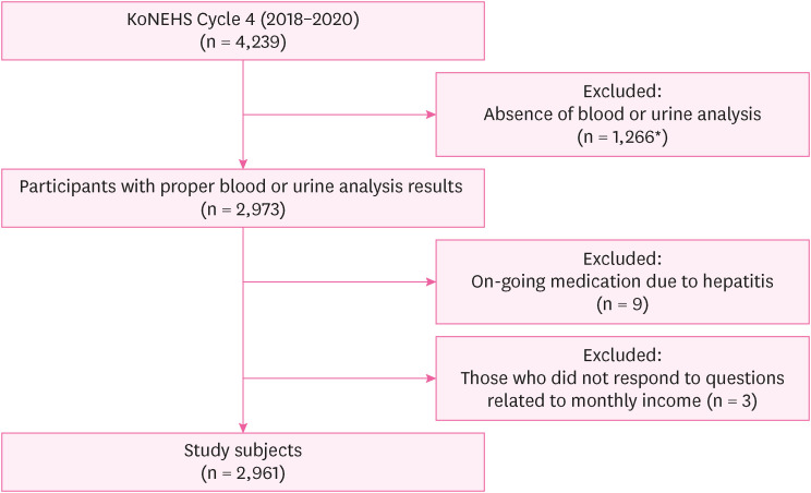
Flow of study participants selection.
KoNEHS: Korean National Environmental Health Survey.
*Due to COVID-19, blood and urine sampling was not properly performed in 2020.
The variables were divided into 2 groups. First, environmentally hazardous chemicals can be identified using blood or urine tests. These substances can be categorized as heavy metals, polyaromatic hydrocarbons, phthalate metabolites, bisphenols, triclosan, parabens, nicotine derivatives, benzophenone, benzene metabolites, perfluorinated compounds, and phenolic benzoic acid. A total of 34 variables were investigated using blood and urine samples. Details are presented in Supplementary Table 1. The second group consisted of questionnaires. The KoNEHS survey contains many questionnaires that can indirectly reflect exposure to various chemicals. The responses to each questionnaire were mostly based on the frequency level, so they could be stratified. A total of 38 questionnaires were identified. The details of the questionnaires are provided in Supplementary Table 2.
We distinguished the liver dysfunction group from the normal population based on elevated liver function indices (aspartate aminotransferase [AST], alanine aminotransferase [ALT], and gamma glutamyl transferase [GGT]). We followed the cut-off values suggested by the KoNEHS Laboratory Procedure Manual. Values greater than AST, 34 IU/L; ALT, 49 IU/L; and GGT, 73 IU/L (for males) or 38 IU/L (for females) were defined as increased liver function indices; therefore, the liver dysfunction group. Urine samples for the measurement of various environmentally hazardous substances were adjusted by urine creatinine concentration. In general, environmental exposure to various substances is low-concentration exposure and shows skewness. Therefore, we log-transformed urine and blood samples for analysis.15 As we mentioned, the questionnaire-based variables are composed of stratified answers based on frequency; we divide the high- and low-exposure groups by binary classification. Few nominal variables were analyzed in the factorization process.
We analyzed the association between various variables of environmental exposure and elevated liver enzyme levels by modifying the EWAS approach proposed by Patel et al.5,16 First, we used a generalized linear model (GLM) to associate the 72 environmental variables with the levels of AST, ALT, and GGT while adjusting for age, sex, smoking, alcohol intake, body mass index (BMI), and monthly income. We calculated odds ratio (OR) of each variable against each liver function indices through GLM, meaning odds of the outcome per unit increase in the value of the exposure. This study has the feature of multiple comparisons using various variables; consequently, there is a possibility of an increase in type I errors. To correct this problem, traditionally, few methods, such as Bonferroni correction, have been used. However, in the case of statistical analyses with a large number of variables, traditional methods are too conservative, resulting in diminished statistical power. Instead, the false discovery rate (FDR) is known to be a less conservative correction method compared to other methods and is widely used in GWAS-based studies.17,18 Therefore, we used the FDR to adjust the p-values when judging the significance of the GLM results. We considered environmentally hazardous substances with FDR < 0.05 as potential risk factors for elevated liver enzyme levels. Previous studies using EWAS analysis used additional validation studies to ensure replicability in other data sets.16,19 However, since the data set we used, KoNEHS, does not have a sufficiently large number of study subjects to properly validate with other sets of studies, we did not perform the additional validation process. Finally, we visualized the association results using a Manhattan plot. All statistical analyses were performed with R software (version 4.2.1 for Windows; R Foundation, Vienna, Austria).
This study was conducted after obtaining approval from the Institutional Review Board (IRB) of Korea University Medical Center (IRB No. 2022AS0310).
RESULTS
A total of 2,961 adults were included in the analysis; 1,289 participants were male (43.5%), and the mean age of the participants was 52.01 (range, 19–82 years). The mean BMI was 25.03 kg/m2. Among the 2,961 participants, 370 had elevated AST (12.5%), 195 had elevated ALT (6.6%), and 342 had elevated GGT (11.6%). In general, the elevation of liver function indices was more common in males than in females. Additionally, liver dysfunction was more prevalent in the overweight and obese groups. The heavy drinking (take alcohol more than 1–2 times a week) and current smoking groups also had higher levels of AST, ALT, and GGT. Elevation of AST levels was observed in the high-income group, but there were no significant differences in ALT and GGT levels (Table 1).
Table 1
General characteristics of subjects
Values are presented as number (%) or mean ± standard deviation.
Monthly income (average monthly household income over the past year): Group 1, less than 3 million won; Group 2, equal or more than 3 million won.
Drinking: None, people who answered they do not drink; Group 1, people who drinks less than once in a month; Group 2, people who drinks 1–2 times in a month; Group 3, people who drinks 1–2 times in a week; Group 4, people who drinks more than 3 times in a week; Group 5, people who drinks almost everyday.
BMI: body mass index; AST: aspartate aminotransferase; ALT: alanine aminotransferase; GGT: gamma glutamyl transferase.
*p < 0.05; **p < 0.01; ***p < 0.001.
Table 2 shows the significant findings of the EWAS study, with an FDR < 0.05. Perfluorooctanesulfonate (PFOS) and perfluorodecanoic acid (PFDeA) were positively associated with liver function indices. PFOS was positively associated with ALT levels (OR: 2.2; 95% confidence interval [CI]: 1.39–3.46; p
adjusted = 0.0147). Furthermore, PFDeA and GGT were positively associated with GGT (OR: 2.73; 95% CI: 1.36–5.5; p
adjusted = 0.0256). In the heavy metals category, high plasma mercury levels were significantly associated with higher GGT levels (OR: 1.45; 95% CI: 1.14–1.84; p
adjusted = 0.0315). Using a plastic container while keeping food in the refrigerator was associated with elevated GGT compared to using a glass container (OR: 1.51; 95% CI: 1.16–1.95; p
adjusted = 0.0153). 2-ethyl-5-oxohexyl phthalate (MEOHP), one of the phthalate metabolites, showed a negative trend with all 3 indices, with AST (OR: 0.54; 95% CI: 0.39–0.73; p
adjusted = 0.00357), ALT (OR: 0.5; 95% CI: 0.34–0.75; p
adjusted = 0.036), GGT (OR: 0.55; 95% CI: 0.4–0.76; p
adjusted = 0.00697). Bisphenol S (BPS), one of the substitute materials for bisphenol A (BPA), showed a negative association with ALT (OR: 0.77; 95% CI: 0.66–0.89; p
adjusted = 0.0151). Frequent use of sunblock cream and level of GGT showed a negative association (OR: 0.25; 95% CI: 0.11–0.55; p
adjusted = 0.036). The frequent use of sunblock cream group is defined as a group that applies sunblock cream more than once or twice a week. Finally, using the Manhattan plot for EWAS analysis, we identified that one factor, 3 factors, and 5 factors among the 72 environmental exposure variables showed statistically significant associations with AST, ALT, and GGT levels, respectively (Fig. 2).
Table 2
Summary of statistically significant associations with abnormal liver function group
Among 72 variables, one variable for AST, 3 variables for ALT, 5 variables for GGT were identified as statistically significant association. To correct possible type I error, p-values were adjusted with the methodology of FDR. Cut-off value of statistically significant result was FDR < 0.05.
OR: odds ratio; CI: confidence interval; FDR: false discovery rate; AST: aspartate aminotransferase; ALT: alanine aminotransferase; GGT: gamma glutamyl transferase; MEOHP: 2-ethyl-5-oxohexyl phthalate; BPS: bisphenol S; PFOS: perfluorooctanesulfonate; PFDeA: perfluorodecanoic acid.
*FDR < 0.05; **FDR < 0.01.
Fig. 2
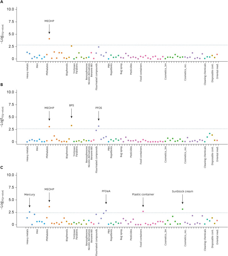
Manhattan plot of EWAS of environmentally hazardous chemicals for elevated liver enzymes. (A) Manhattan plot of EWAS for elevated aspartate aminotransferase. (B) Manhattan plot of EWAS for elevated alanine aminotransferase. (C) Manhattan plot of EWAS for elevated gamma glutamyl transferase. X-axis shows the groups of categorized chemicals. Y-axis shows −log10(p-value) of the generalized linear model analysis with each variables with elevated liver enzymes. Each categorized group was represented as same color of dots. The statistically significant cut-off values of false discovery rate < 0.05 were represented as grey dashed line.
MEOHP: 2-ethyl-5-oxohexyl phthalate; PAH: polycyclic aromatic hydrocarbon; PBA: 3-phenoxybenzoic acid; BPS: bisphenol S; PFOS: perfluorooctanesulfonate; PFDeA: perfluorodecanoic acid; EWAS: environment-wide association study; 3m: 3 months; rec.: recent; cont.: container; med.: medicine.
DISCUSSION
This exploratory study aimed to identify the relationship between multiple exposures to chemical agents and human liver function. As mentioned earlier, exposure to chemicals in our daily life has the characteristics of chronic, low-concentration, simultaneous concurrent complicated exposure.3,4 In this study, we borrowed the idea of the EWAS methodology to reflect these exposure characteristics. As a result, we identified 7 factors that could potentially influence liver function. Among these factors, 4 were identified as potential risk factors for the elevation of liver inflammatory indices.
Since this study is exploratory, matching and confirming our findings with other studies is crucial. Some factors showed a similar trend in association with previous studies; however, other factors showed different results. Plasma mercury level was positively associated with GGT level (OR: 1.45; 95% CI: 1.14–1.84; p
adjusted = 0.0315). This trend has been confirmed in many other studies. Seo et al.20 investigated 1,959 Korean subjects using data from the 2019 Korean National Health and Nutrition Examination Survey and found a positive association between blood mercury concentration and GGT level. In some studies, a high blood mercury level is associated with higher AST and ALT, which means that mercury could cause an overall inflammatory burden on the liver.21 This elevation of liver inflammatory indices can be explained by mechanisms of oxidative stress, disruption of metabolism, and cell death. Exposure to mercury suppresses various antioxidant mechanisms by amplifying free radical production, resulting in increased protective enzymes such as GGT.22,23 In addition to oxidative stress in the liver, mercury can potentially interrupt various endocrine mechanisms throughout the human body. A previous study by Hu et al.24 reported a positive association between mercury and blood pressure. He et al.25 conducted a prospective cohort study of 3,875 young adults in the US and found that high exposure to mercury in young adulthood may increase the risk of diabetes later in life. In general, mercury exposure is mostly due to organic dietary intake,26,27 and most environmental exposure remains below the subclinical level, yet various health effects of mercury need to be identified.
In the perfluorinated compound category, PFOS showed a positive association with ALT (OR: 2.2; 95% CI: 1.39–3.46; p
adjusted = 0.0147), and PFDeA also showed a positive association with GGT (OR: 2.73; 95% CI: 1.36–5.5; p
adjusted = 0.0256). Perfluorinated compounds are widely used in industrial and household applications; they are used in various parts of human life, including coating materials for cookware, waterproof and stain-resistant fabrics, various packaging materials, and firefighting materials.28 Due to its universal use in daily life, its potential health effects on human body have attracted attention. As a result, many studies have been performed on perfluorinated compounds. According to a systematic review and meta-analysis by Costello et al.,29 which yielded 85 rodent studies and 24 epidemiological studies of humans, the legacy of PFAS showed liver toxicity in humans and rodents. This study showed a positive association between ALT and exposure to various perfluorinated compounds, including perfluorooctanoic acid (PFOA) and PFOS. In animal studies, PFAS showed a disruption of lipid metabolism, resulting in consistent accumulation of lipids and induction of steatosis.30,31 It is also known that PFOS and PFOA, subfamilies of PFAS, can cause liver enlargement in rodents and hepatocellular adenomas in rats.32 The peroxisome proliferator-activated receptor-alpha, a major regulator of lipid metabolism in the liver, plays a role in these metabolic disruptions.33,34,35 People who used plastic containers for food storage in refrigerators showed higher levels of GGT than those who used glass containers for storage (OR: 1.51; 95% CI: 1.16–1.95; p
adjusted = 0.0153). Studies on the direct association between the use of plastic containers and human liver function are difficult to find. However, plastic containers usually contain hazardous substances such as bisphenols, phthalate metabolites, polyvinyl chloride, and perfluorinated alkylated substances.36,37 Since these chemicals are proven to be hepatotoxic, the use of plastic containers in daily life could affect liver function in this manner.
MEOHP, a phthalate metabolite, showed a negative association with AST (OR: 0.54; 95% CI: 0.39–0.73; p
adjusted = 0.00357), ALT (OR: 0.5; 95% CI: 0.34–0.75; p
adjusted = 0.036), and GGT (OR: 0.55; 95% CI: 0.4–0.76; p
adjusted = 0.00697). Phthalates are widely used as plasticizers and additives in cosmetics.38 Since phthalates are used in almost all modern industries, studies to determine their health effects on humans have been widely performed. Phthalates are well known for their endocrine-disrupting mechanisms, including suppression of the reproductive tract, childhood obesity, early menarche,38,39 and other adverse effects in various organs are actively revealed. MEOHP is a metabolite of di-2-ethylhexyl phthalate (DEHP). In general, DEHP has various adverse effects on multiple organs, including the liver.40 The negative association of MEOHP and liver function indices in this study could be thought to have inconsistent outcomes compared to previous studies. However, some studies showed similar trends in the relationship between GGT and MEOHP.41 MEOHP is a relatively stable form of DEHP derivatives. Among the metabolites of DEHP, mono (2-ethylhexyl) phthalate (MEHP), which can be converted to MEOHP through hydroxylation and oxidation, has the most bioactive traits.42 Therefore, higher levels of MEOHP could indicate that an individual’s ability to convert more bioactive substances (MEHP) to relatively less toxic substances (MEOHP) is high, resulting in a lower inflammatory index of the liver (GGT).41,43,44
BPS showed a negative association with the level of ALT (OR: 0.77; 95% CI: 0.66–0.89; p
adjusted = 0.0151). As BPA is well known for its toxic effects on human health, BPS was introduced as a substitute. However, many in vitro studies have recently shown that BPS has toxic effects on various organs, including the liver.45 Our study showed different trends from these studies; we could not find solid evidence that could support our results. This might be because there are always possibilities of inconsistency in the trends of the results due to differences in study design or populations or methods to evaluate the level of exposure.41 Therefore, more studies are crucial. The frequency of sunblock cream use over the last 3 months showed a negative relationship with the level of GGT (OR: 0.25; 95% CI: 0.11–0.55; p
adjusted = 0.036). Studies investigating the association between sunblock cream use and human liver function are not readily available. There has been a report that the use of sunscreens under sun radiation shows elevated ALT and AST in the animal study.46 In general, sunlight is believed to have anti-inflammatory effects on the liver by the production of active vitamin D (1,25(OH)2D).47 This trend could be partially inconsistent with our results. However, in our study, we defined the high-exposure group to sunblock cream based on a questionnaire asking, “How often did you wear sunblock cream for the last 3 months?” We could not quantify the actual use of chemicals or the actual amount of sunlight that is exposed by these questions. This could be why our result was somewhat inconsistent with previous literature and, at the same time, could be one of the limitations of this study.
One of the strengths of this study, in EWAS, is that we could investigate global and comprehensive patterns of association between various factors and certain diseases. Traditional epidemiologic approaches to diseases have focused on the relationship between specific factors or a very limited number of factors and diseases. The EWAS methodology could provide a global view of these associations.16 Furthermore, visualization of the result with a Manhattan plot could strengthen this study by comprehensively skimming the relationships between factors. For example, in Fig. 2C, the p-values of the heavy metals and fluorinated compounds are distributed near the set point of FDR of 0.05. These variables did not reach the cut-off value of statistical significance (FDR < 0.05), but we still have an overall understanding that the components of these groups tend to have a higher association with liver function.
This study has some limitations. First, since this was a cross-sectional study, the causal inference of the results would be based on an assumption. Second, many variables in this study were based on questionnaires. We tried to indirectly determine individuals’ life patterns or habits, implying their exposure to chemicals. Questionnaires are the only way to evaluate the life patterns and habits of individuals. However, questionnaires have the possibility of recall bias. Third, again from the point of questionnaires, although we designed the variables into a binary classification based on frequency, the responses to the questionnaires cannot exactly reflect the actual amount of exposure. Therefore, there is the possibility of an unclear or partial disruption in the analysis. EWAS studies usually require a large number of variables, our study included 72 variables, of which only 34 were data from blood and urine analyses. This is a relatively low number of variables compared to large data sets such as the National Health and Nutrition Examination Survey in the US. However, we attempted to perform an exploratory analysis based on the Korean population; therefore, the KoNEHS was the best survey data we could use. Finally, as mentioned earlier, our dataset does not contain a sufficient number of study subjects to perform a validation process. The absence of a validation step could show limitations in securing the replicability of the analysis.
CONCLUSIONS
In conclusion, we applied the EWAS methodology to investigate the association of various environmentally hazardous chemicals with human liver function in the Korean population using the 4th KoNEHS data. Seven potential risk factors were identified. Exploratory studies on various diseases can be carried out by applying the EWAS frame.
ACKNOWLEDGEMENTS
We appreciate National Institute of Environmental Research making available the raw data of Korean National Environmental Health Survey.
Abbreviations
AST
aspartate aminotransferase
ALT
alanine aminotransferase
BMI
body mass index
BPA
bisphenol A
BPS
bisphenol S
CI
confidence interval
cont.
container
DEHP
di-2-ethylhexyl phthalate
EWAS
environment-wide association study
FDR
false discovery rate
GGT
gamma glutamyl transferase
GLM
generalized linear model
GWAS
genome-wide association study
IRB
Institutional Review Board
KoNEHS
Korean National Environmental Health Survey
MEHP
mono (2-ethylhexyl) phthalate
MEOHP
2-ethyl-5-oxohexyl phthalate
med.
medicine
OR
odds ratio
PAH
polycyclic aromatic hydrocarbon
PBA
3-phenoxybenzoic acid
PFAS
polyfluorinated substances
PFDeA
perfluorodecanoic acid
PFOA
perfluorooctanoic acid
PFOS
perfluorooctanesulfonate
rec.
recent
3m
three months
-
Funding: This research was supported by the Inha University Hospital’s Environmental Health Center for Training Environmental Medicine Professionals funded by the Ministry of Environment, Republic of Korea (2022).
-
Competing interests: The authors declare that they have no competing interests.
-
Author Contributions:
NOTES
SUPPLEMENTARY MATERIALS
Supplementary Table 1
List of environmentally hazardous substances analyzed with blood and urine samples
- 1. European Union. The European environment – state and outlook 2020. Updated 2020]. Accessed December 15, 2022]. http://www.eea.europa.eu/soer/publications/soer-2020 .
- 2. World Health Organization. Preventing noncommunicable diseases (NCDs) by reducing environmental risk factors. Updated 2017]. Accessed December 15, 2022]. https://apps.who.int/iris/bitstream/handle/10665/258796/WHO-FWC-EPE-17.01-eng.pdf .
- 3. Kortenkamp A, Faust M, Scholze M, Backhaus T. Low-level exposure to multiple chemicals: reason for human health concerns? Environ Health Perspect 2007;115:Suppl 1. (Suppl 1):106–114. 18174958.ArticlePubMedPMC
- 4. Sprinkle RH, Payne-Sturges DC. Mixture toxicity, cumulative risk, and environmental justice in United States federal policy, 1980-2016: why, with much known, was little done? Environ Health 2021;20(1):104. 34535123.PubMedPMC
- 5. Patel CJ. Introduction to environment and exposome-wide association studies: a data-driven method to identify multiple environmental factors associated with phenotypes in human populations. In: Rider CV, Simmons JE, editors. Chemical Mixtures and Combined Chemical and Nonchemical Stressors. Cham, Switzerland: Springer International Publishing AG; 2018, 129–149.
- 6. Rappaport SM, Barupal DK, Wishart D, Vineis P, Scalbert A. The blood exposome and its role in discovering causes of disease. Environ Health Perspect 2014;122(8):769–774. 24659601.ArticlePubMedPMC
- 7. Wahlang B, Beier JI, Clair HB, Bellis-Jones HJ, Falkner KC, McClain CJ, et al. Toxicant-associated steatohepatitis. Toxicol Pathol 2013;41(2):343–360. 23262638.ArticlePubMedPMCPDF
- 8. Wahlang B, Jin J, Beier JI, Hardesty JE, Daly EF, Schnegelberger RD, et al. Mechanisms of environmental contributions to fatty liver disease. Curr Environ Health Rep 2019;6(3):80–94. 31134516.ArticlePubMedPMCPDF
- 9. Ledda C, Loreto C, Zammit C, Marconi A, Fago L, Matera S, et al. Non-infective occupational risk factors for hepatocellular carcinoma: a review (review). Mol Med Rep 2017;15(2):511–533. 28000892.PubMed
- 10. National Center for Environmental Health. Fourth national report on human exposure to environmental chemicals. Updated tables, March 2021: volume three: analysis of pooled serum samples for select chemicals, NHANES 2005-2016. Updated 2021]. Accessed December 15, 2022]. https://stacks.cdc.gov/view/cdc/105344 .
- 11. Li Y, Fletcher T, Mucs D, Scott K, Lindh CH, Tallving P, et al. Half-lives of PFOS, PFHxS and PFOA after end of exposure to contaminated drinking water. Occup Environ Med 2018;75(1):46–51. 29133598.ArticlePubMedPMC
- 12. Zhao M, Ge X, Xu J, Li A, Mei Y, Yin G, et al. Association between urine metals and liver function biomarkers in Northeast China: a cross-sectional study. Ecotoxicol Environ Saf 2022;231:113163. 35030523.ArticlePubMed
- 13. Chang WJ, Joe KT, Park HY, Jeong JD, Lee DH. The relationship of liver function tests to mixed exposure to lead and organic solvents. Ann Occup Environ Med 2013;25(1):5. 24472152.ArticlePubMedPMC
- 14. Cave M, Appana S, Patel M, Falkner KC, McClain CJ, Brock G. Polychlorinated biphenyls, lead, and mercury are associated with liver disease in American adults: NHANES 2003-2004. Environ Health Perspect 2010;118(12):1735–1742. 21126940.ArticlePubMedPMC
- 15. Feng C, Wang H, Lu N, Chen T, He H, Lu Y, et al. Log-transformation and its implications for data analysis. Shanghai Arch Psychiatry 2014;26(2):105–109. 25092958.PubMedPMC
- 16. Patel CJ, Bhattacharya J, Butte AJ. An environment-wide association study (EWAS) on type 2 diabetes mellitus. PLoS One 2010;5(5):e10746. 20505766.ArticlePubMedPMC
- 17. Storey JD, Tibshirani R. Statistical significance for genomewide studies. Proc Natl Acad Sci U S A 2003;100(16):9440–9445. 12883005.ArticlePubMedPMC
- 18. Benjamini Y, Cohen R. Weighted false discovery rate controlling procedures for clinical trials. Biostatistics 2017;18(1):91–104. 27445132.ArticlePubMedPMC
- 19. Hall MA, Dudek SM, Goodloe R, Crawford DC, Pendergrass SA, Peissig P, et al. Environment-wide association study (EWAS) for type 2 diabetes in the Marshfield Personalized Medicine Research Project Biobank. Pac Symp Biocomput 2014;200–211.Article
- 20. Seo MS, Lee HR, Shim JY, Kang HT, Lee YJ. Relationship between blood mercury concentrations and serum γ-glutamyltranspeptidase level in Korean adults using data from the 2010 Korean National Health and Nutrition Examination Survey. Clin Chim Acta 2014;430:160–163. 24508988.ArticlePubMed
- 21. Choi J, Bae S, Lim H, Lim JA, Lee YH, Ha M, et al. Mercury exposure in association with decrease of liver function in adults: a longitudinal study. J Prev Med Public Health 2017;50(6):377–385. 29207447.ArticlePubMedPMCPDF
- 22. Lim JS, Yang JH, Chun BY, Kam S, Jacobs DR Jr, Lee DH. Is serum gamma-glutamyltransferase inversely associated with serum antioxidants as a marker of oxidative stress? Free Radic Biol Med 2004;37(7):1018–1023. 15336318.PubMed
- 23. Farina M, Avila DS, da Rocha JB, Aschner M. Metals, oxidative stress and neurodegeneration: a focus on iron, manganese and mercury. Neurochem Int 2013;62(5):575–594. 23266600.ArticlePubMedPMC
- 24. Hu XF, Singh K, Chan HM. Mercury exposure, blood pressure, and hypertension: a systematic review and dose-response meta-analysis. Environ Health Perspect 2018;126(7):076002. 30073953.ArticlePubMedPMC
- 25. He K, Xun P, Liu K, Morris S, Reis J, Guallar E. Mercury exposure in young adulthood and incidence of diabetes later in life: the CARDIA Trace Element Study. Diabetes Care 2013;36(6):1584–1589. 23423697.PubMedPMC
- 26. Mahaffey KR, Mergler D. Blood levels of total and organic mercury in residents of the upper St. Lawrence River basin, Québec: association with age, gender, and fish consumption. Environ Res 1998;77(2):104–114. 9600803.ArticlePubMed
- 27. Mozaffarian D, Rimm EB. Fish intake, contaminants, and human health: evaluating the risks and the benefits. JAMA 2006;296(15):1885–1899. 17047219.ArticlePubMed
- 28. Buck RC, Franklin J, Berger U, Conder JM, Cousins IT, de Voogt P, et al. Perfluoroalkyl and polyfluoroalkyl substances in the environment: terminology, classification, and origins. Integr Environ Assess Manag 2011;7(4):513–541. 21793199.ArticlePubMedPMCPDF
- 29. Costello E, Rock S, Stratakis N, Eckel SP, Walker DI, Valvi D, et al. Exposure to per- and polyfluoroalkyl substances and markers of liver injury: a systematic review and meta-analysis. Environ Health Perspect 2022;130(4):46001. 35475652.ArticlePubMedPMC
- 30. Das KP, Wood CR, Lin MT, Starkov AA, Lau C, Wallace KB, et al. Perfluoroalkyl acids-induced liver steatosis: effects on genes controlling lipid homeostasis. Toxicology 2017;378:37–52. 28049043.ArticlePubMedPMC
- 31. Bagley BD, Chang SC, Ehresman DJ, Eveland A, Zitzow JD, Parker GA, et al. Perfluorooctane sulfonate-induced hepatic steatosis in male Sprague Dawley rats is not attenuated by dietary choline supplementation. Toxicol Sci 2017;160(2):284–298. 28973659.ArticlePubMed
- 32. Lau C, Anitole K, Hodes C, Lai D, Pfahles-Hutchens A, Seed J. Perfluoroalkyl acids: a review of monitoring and toxicological findings. Toxicol Sci 2007;99(2):366–394. 17519394.ArticlePubMed
- 33. Abdellatif A, Al-Tonsy AH, Awad ME, Roberfroid M, Khan MN. Peroxisomal enzymes and 8-hydroxydeoxyguanosine in rat liver treated with perfluorooctanoic acid. Dis Markers 2003-2004;19(1):19–25.ArticlePubMedPMCPDF
- 34. Bjork JA, Butenhoff JL, Wallace KB. Multiplicity of nuclear receptor activation by PFOA and PFOS in primary human and rodent hepatocytes. Toxicology 2011;288(1-3):8–17. 21723365.ArticlePubMed
- 35. Elcombe CR, Elcombe BM, Foster JR, Farrar DG, Jung R, Chang SC, et al. Hepatocellular hypertrophy and cell proliferation in Sprague-Dawley rats following dietary exposure to ammonium perfluorooctanoate occurs through increased activation of the xenosensor nuclear receptors PPARα and CAR/PXR. Arch Toxicol 2010;84(10):787–798. 20614104.ArticlePubMedPDF
- 36. Vered G, Shenkar N. Monitoring plastic pollution in the oceans. Curr Opin Toxicol 2021;27:60–68.Article
- 37. Proshad R, Kormoker T, Islam S, Haque MA, Rhaman M, Mithu MR. Toxic effects of plastic on human health and environment: aconsequences of health risk assessment in Bangladesh. Int J Heal 2018;6(1):1–5.ArticlePDF
- 38. Katsikantami I, Sifakis S, Tzatzarakis MN, Vakonaki E, Kalantzi OI, Tsatsakis AM, et al. A global assessment of phthalates burden and related links to health effects. Environ Int 2016;97(97):212–236. 27669632.ArticlePubMed
- 39. Park O, Park JT, Chi Y, Kwak K. Association of phthalates and early menarche in Korean adolescent girls from Korean National Environmental Health Survey (KoNEHS) 2015-2017. Ann Occup Environ Med 2021;33(1):e4. 34754465.ArticlePubMedPMCPDF
- 40. Rowdhwal SS, Chen J. Toxic effects of di-2-ethylhexyl phthalate: an overview. BioMed Res Int 2018;2018:1750368. 29682520.ArticlePubMedPMCPDF
- 41. Ferguson KK, Loch-Caruso R, Meeker JD. Urinary phthalate metabolites in relation to biomarkers of inflammation and oxidative stress: NHANES 1999-2006. Environ Res 2011;111(5):718–726. 21349512.ArticlePubMedPMC
- 42. Erkekoglu P, Rachidi W, Yuzugullu OG, Giray B, Favier A, Ozturk M, et al. Evaluation of cytotoxicity and oxidative DNA damaging effects of di(2-ethylhexyl)-phthalate (DEHP) and mono(2-ethylhexyl)-phthalate (MEHP) on MA-10 Leydig cells and protection by selenium. Toxicol Appl Pharmacol 2010;248(1):52–62. 20659492.ArticlePubMed
- 43. Hauser R. Urinary phthalate metabolites and semen quality: a review of a potential biomarker of susceptibility. Int J Androl 2008;31(2):112–117. 18067563.ArticlePubMed
- 44. Meeker JD, Calafat AM, Hauser R. Di(2-ethylhexyl) phthalate metabolites may alter thyroid hormone levels in men. Environ Health Perspect 2007;115(7):1029–1034. 17637918.ArticlePubMedPMC
- 45. Qin J, Ru S, Wang W, Hao L, Ru Y, Wang J, et al. Long-term bisphenol S exposure aggravates non-alcoholic fatty liver by regulating lipid metabolism and inducing endoplasmic reticulum stress response with activation of unfolded protein response in male zebrafish. Environ Pollut 2020;263(Pt B):114535. 32283406.ArticlePubMed
- 46. Al-Eitan LN, Aljamal HA, Alkhatib RQ. Gas chromatographic-mass spectrometric analysis of sunscreens and their effects on mice liver and kidney enzyme function. Clin Cosmet Investig Dermatol 2018;12:11–21.PubMedPMC
- 47. Gorman S, Black LJ, Feelisch M, Hart PH, Weller R. Can skin exposure to sunlight prevent liver inflammation? Nutrients 2015;7(5):3219–3239. 25951129.ArticlePubMedPMC
REFERENCES
REFERENCES
Figure & Data
REFERENCES
Citations
Citations to this article as recorded by 
- Metals exposure and biomarkers of liver damage: a systematic review and meta-analysis of observational studies
Ibrahim Issah, Serwaa A. Bawua, John Arko-Mensah, Mabel S. Duah, Shirley V. Simpson, Thomas P. Agyekum, Olalekan A. Uthman, Julius N. Fobil
Reviews on Environmental Health.2026; 41(1): 109. CrossRef - Association between gut microbiota composition and liver dysfunction in school-aged children exposed to triclosan
Aijing Li, Menglong Li, Wang Xu, Yanhui Zhang, Shulan Yin, Yating Yu, Shufa Zheng, Yifei Hu, Maoyong Song
Journal of Environmental Sciences.2026; 167: 308. CrossRef - Exposome-wide association study of cognitive function in US older adults using the NHANES data
Hyuna Jang, Jiyun Lee, Vy Kim Nguyen, Hyeong-Moo Shin
Exposome.2025;[Epub] CrossRef - Liver Toxicity Induced by Exposure to Bisphenol Analogs at Environmentally Relevant Levels: Insights from a Literature Review on Multiple Species
Tai L. Guo, Fatma Eldefrawy, Kevin M. Guo
Livers.2025; 5(2): 24. CrossRef - Sex Differences in the Association Between the Korean Healthy Eating Index and Liver Enzymes Among Korean Adults
Seong-Uk Baek, Jin-Ha Yoon
Nutrients.2025; 17(14): 2372. CrossRef - Association Between Heavy Metals Exposure and Elevated High-Sensitivity C-Reactive Protein: Mediating Role of Body Mass Index
Seong-Uk Baek, Jin-Ha Yoon
Biomolecules.2025; 15(11): 1491. CrossRef
 Cite
Cite- Figure
-
- Related articles
-
- Occupational differences in benzene-related biomarker levels beyond traditional industrial settings: findings from the Korean National Environmental Health Survey, 2015–2023
- Relationship between the use of hair products and urine benzophenone-3: the Korean National Environmental Health Survey (KoNEHS) cycle 4
Environment-wide association study of elevated liver enzymes: results from the Korean National Environmental Health Survey 2018–2022


Fig. 1 Flow of study participants selection.KoNEHS: Korean National Environmental Health Survey.*Due to COVID-19, blood and urine sampling was not properly performed in 2020.
Fig. 2 Manhattan plot of EWAS of environmentally hazardous chemicals for elevated liver enzymes. (A) Manhattan plot of EWAS for elevated aspartate aminotransferase. (B) Manhattan plot of EWAS for elevated alanine aminotransferase. (C) Manhattan plot of EWAS for elevated gamma glutamyl transferase. X-axis shows the groups of categorized chemicals. Y-axis shows −log10(p-value) of the generalized linear model analysis with each variables with elevated liver enzymes. Each categorized group was represented as same color of dots. The statistically significant cut-off values of false discovery rate < 0.05 were represented as grey dashed line.MEOHP: 2-ethyl-5-oxohexyl phthalate; PAH: polycyclic aromatic hydrocarbon; PBA: 3-phenoxybenzoic acid; BPS: bisphenol S; PFOS: perfluorooctanesulfonate; PFDeA: perfluorodecanoic acid; EWAS: environment-wide association study; 3m: 3 months; rec.: recent; cont.: container; med.: medicine.
Fig. 1
Fig. 2
Environment-wide association study of elevated liver enzymes: results from the Korean National Environmental Health Survey 2018–2022
| Characteristics | All (n = 2,961) | Dysfunction group | ||||||
|---|---|---|---|---|---|---|---|---|
| AST (n = 370) | ALT (n = 195) | GGT (n = 342) | ||||||
| Sex | < 0.001*** | < 0.001*** | < 0.01** | |||||
| Male | 1,289 (43.5) | 215 (58.1) | 134 (68.7) | 175 (51.2) | ||||
| Female | 1,672 (56.5) | 155 (41.9) | 61 (31.3) | 167 (48.8) | ||||
| Age | 52.01 ± 14.0 | 56.1 ± 13.6 | < 0.001*** | 47.8 ± 14.9 | < 0.001*** | 53.8 ± 12.8 | 0.092 | |
| BMI | < 0.001*** | < 0.001*** | < 0.001*** | |||||
| < 23 | 872 (29.5) | 67 (18.1) | 10 (5.1) | 60 (17.5) | ||||
| 23–25 | 679 (22.9) | 67 (18.1) | 32 (16.4) | 68 (19.9) | ||||
| ≥ 25 | 1,410 (47.6) | 236 (63.8) | 153 (78.5) | 214 (62.6) | ||||
| Drinking | < 0.001*** | < 0.05* | < 0.001*** | |||||
| None | 912 (30.8) | 109 (29.5) | 46 (23.6) | 72 (21.1) | ||||
| Group 1 | 473 (16.0) | 46 (12.4) | 23 (11.8) | 38 (11.1) | ||||
| Group 2 | 524 (17.7) | 52 (14.1) | 38 (19.5) | 35 (10.2) | ||||
| Group 3 | 588 (19.9) | 76 (20.5) | 48 (24.6) | 70 (20.5) | ||||
| Group 4 | 301 (10,2) | 53 (14.3) | 26 (13.3) | 72 (21.1) | ||||
| Group 5 | 163 (5.5) | 34 (9.2) | 14 (7.2) | 55 (16.1) | ||||
| Smoking | < 0.001*** | < 0.001*** | < 0.001*** | |||||
| Never | 1,905 (64.3) | 193 (52.2) | 94 (48.2) | 189 (55.3) | ||||
| Quit | 593 (20.0) | 101 (27.3) | 44 (22.6) | 66 (19.3) | ||||
| Current | 463 (15.7) | 76 (20.5) | 57 (29.2) | 87 (25.4) | ||||
| Monthly income | < 0.001*** | 0.301 | 0.097 | |||||
| Group 1 | 1,343 (45.4) | 208 (56.2) | 81 (41.5) | 170 (49.7) | ||||
| Group 2 | 1,618 (54.6) | 162 (43.8) | 114 (58.5) | 172 (50.3) | ||||
| Hazardous substances | OR (95% CI) | FDR | |||
|---|---|---|---|---|---|
| Abnormal AST | |||||
| Phthalates | |||||
| MEOHP (µg/L) | 0.54 (0.39–0.73) | 8.40 × 10⁻⁵ | 3.57 × 10⁻³** | ||
| Abnormal ALT | |||||
| Phthalates | |||||
| MEOHP (µg/L) | 0.5 (0.34–0.75) | 8.46 × 10⁻⁴ | 3.60 × 10⁻²* | ||
| Bisphenols | |||||
| BPS (µg/L) | 0.77 (0.66–0.89) | 5.32 × 10⁻⁴ | 1.51 × 10⁻²* | ||
| Perfluorinated compounds | |||||
| PFOS (µg/L) | 2.2 (1.39–3.46) | 6.93 × 10⁻⁴ | 1.47 × 10⁻²* | ||
| Abnormal GGT | |||||
| Heavy metals | |||||
| Mercury (plasma) (µg/L) | 1.45 (1.14–1.84) | 2.59 × 10⁻³ | 3.15 × 10⁻²* | ||
| Phthalates | |||||
| MEOHP (µg/L) | 0.55 (0.4–0.76) | 2.46 × 10⁻⁴ | 6.97 × 10⁻³** | ||
| Perfluorinated compounds | |||||
| PFDeA (µg/L) | 2.73 (1.36–5.5) | 1.50 × 10⁻³ | 2.56 × 10⁻²* | ||
| Food container | |||||
| Plastic (compared to glass) | 1.51 (1.16–1.95) | 2.13 × 10⁻³ | 1.53 × 10⁻²* | ||
| Cosmetics | |||||
| Wearing sun block cream (≥ 1–2 times per week) | 0.25 (0.11–0.55) | 7.18 × 10⁻⁴ | 3.02 × 10⁻²* | ||
Table 1 General characteristics of subjects
Values are presented as number (%) or mean ± standard deviation.
Monthly income (average monthly household income over the past year): Group 1, less than 3 million won; Group 2, equal or more than 3 million won.
Drinking: None, people who answered they do not drink; Group 1, people who drinks less than once in a month; Group 2, people who drinks 1–2 times in a month; Group 3, people who drinks 1–2 times in a week; Group 4, people who drinks more than 3 times in a week; Group 5, people who drinks almost everyday.
BMI: body mass index; AST: aspartate aminotransferase; ALT: alanine aminotransferase; GGT: gamma glutamyl transferase.
*
Table 2 Summary of statistically significant associations with abnormal liver function group
Among 72 variables, one variable for AST, 3 variables for ALT, 5 variables for GGT were identified as statistically significant association. To correct possible type I error,
OR: odds ratio; CI: confidence interval; FDR: false discovery rate; AST: aspartate aminotransferase; ALT: alanine aminotransferase; GGT: gamma glutamyl transferase; MEOHP: 2-ethyl-5-oxohexyl phthalate; BPS: bisphenol S; PFOS: perfluorooctanesulfonate; PFDeA: perfluorodecanoic acid.
*FDR < 0.05; **FDR < 0.01.

