 KSOEM
KSOEM

Articles
- Page Path
- HOME > Ann Occup Environ Med > Volume 38; 2026 > Article
- Case Report Reactive airways dysfunction syndrome following acute lithium hydroxide dust exposure at a battery material production plant: a case series of 16 workers
-
Chul Gab Lee1,2,*
 , Soo Hyeong Park1,2, Jeong Joon Park1,2, Han Soo Song1,2, Hyeon Kyeong Ko2, Sung Ho Yoon3
 -
Annals of Occupational and Environmental Medicine 2026;38:e17.
DOI: https://doi.org/10.35371/aoem.2026.38.e17
Published online: June 4, 2026
1Department of Occupational and Environmental Medicine, Chosun University Hospital, Gwangju, Korea
2Gwangju Branch of Korea Occupational Disease Surveillance Center, Gwangju, Korea
3Division of Pulmonology, Department of Internal Medicine, Chosun University Hospital, Gwangju, Korea
- *Corresponding author: Chul Gab Lee Department of Occupational and Environmental Medicine, Chosun University Hospital, 365 Pilmun-daero, Dong-gu, Gwangju 61453, Korea E-mail: eecg@daum.net, cglee@chosun.ac.kr
• Received: April 12, 2026 • Revised: May 29, 2026 • Accepted: May 29, 2026
© 2026 Korean Society of Occupational & Environmental Medicine
This is an Open Access article distributed under the terms of the Creative Commons Attribution Non-Commercial License (https://creativecommons.org/licenses/by-nc/4.0/) which permits unrestricted non-commercial use, distribution, and reproduction in any medium, provided the original work is properly cited.
- 1,751 Views
- 55 Download


Abstract
-
Background Reactive airways dysfunction syndrome (RADS) is an acute-onset form of irritant-induced asthma that occurs after a single high-concentration exposure to irritants. Although RADS has been documented for chlorine, ammonia, and acid fumes, no published case series has attributed RADS to lithium hydroxide (LiOH) dust inhalation. We report the clinical features, diagnostic evaluation, and longitudinal management of 16 workers with persistent respiratory symptoms following acute occupational LiOH exposure at a battery cathode material production facility.
-
Case presentation In March 2024, a silicone connector failure resulted in the leakage of approximately 50–100 kg of LiOH powder at a facility in Korea, exposing over 500 workers. Sixteen workers (15 men, 1 woman; mean age 53.4 years) with persistent respiratory symptoms were evaluated at the Department of Occupational and Environmental Medicine. Most patients presented with cough and sputum production; 56.3% (9/16) exhibited nocturnal or early-morning exacerbation. Methacholine challenge testing was performed in 10 patients, with positive results in three patients (PC20 [provocative concentration causing a 20% decline in forced expiratory volume in 1 second]: 1.36–6.74 mg/mL). By the Brooks 1985 criteria, one case was classified as definite RADS, four as probable, six as possible, and five as unlikely; American College of Chest Physicians 2008 cross-validation yielded identical classifications. Workers' compensation recipients had significantly longer follow-up (18–23 months) compared to non-recipients (1–3 months). Pharmacological management was symptom-directed, combining leukotriene receptor antagonists, mucolytics, antihistamines, and acid-suppressive therapy.
-
Conclusions Acute high-concentration LiOH dust inhalation may induce RADS characterized by persistent respiratory symptoms and bronchial hyperresponsiveness. The extreme alkalinity, high water solubility, and exothermic dissolution of LiOH provide a plausible mechanistic framework for airway injury. Clinicians should consider RADS in workers presenting with persistent respiratory symptoms after alkaline dust exposure, even when routine investigations are unremarkable. Attention should also be given to the psychological burden associated with prolonged, poorly recognized symptoms.
BACKGROUND
The rapid expansion of the lithium-ion battery industry has increased the potential for occupational exposure to lithium hydroxide (LiOH), a highly alkaline compound. Exposure to substantial amounts of LiOH can result in localized respiratory symptoms, such as coughing and sputum production, as well as a variety of systemic symptoms, including headache, nausea, chest tightness, dizziness, and fatigue.1 Although the production processes for LiOH are primarily automated, workers engaged in raw material input, product packaging, and maintenance activities may experience dermatological symptoms, including itching, erythema, and chemical burns.2
In March 2024, during the commissioning of a new LiOH facility in Korea, a silicone connector within the powder supply line sustained damage due to mechanical vibrations. This incident resulted in a leakage of 50 to 100 kg of LiOH powder, impacting hundreds of workers. Reports documented acute health effects.1 Numerous workers exhibited chronic symptoms, including persistent cough, sore throat, foreign body sensation, and shortness of breath, persisting for over four weeks. Despite visits to hospitals in proximity to the factory, no definitive diagnosis or intervention was established, as chest X-rays and blood tests revealed no specific abnormalities. Some workers were suspected of symptom exaggeration or malingering. Consequently, a subset of workers was referred to the occupational and environmental medicine (OEM) outpatient clinic through the Occupational Disease Surveillance Center of Korea (KODSC).
Upon clinical assessment, it was determined that the symptoms in these patients were consistent with reactive airways dysfunction syndrome (RADS), a distinct form of irritant-induced asthma (IIA) first described by Brooks et al. in 1985.3 RADS is characterized by acute asthma-like symptoms after a single high-concentration exposure to irritating vapors, fumes, or dust, without pre-existing respiratory disease, with nonspecific bronchial hyperresponsiveness (BHR) persisting for months to years.3,4 Although RADS has been documented following exposure to chlorine, ammonia, sulfur dioxide, and various acid fumes, there is no published case series specifically associating RADS with LiOH dust inhalation.5,6 The original RADS concept from 1985 involved 10 healthy individuals developing persistent asthma-like symptoms after a single high-concentration irritant exposure,3 and has since evolved. Subsequent observations that did not conform to the single-massive-exposure model led to identifying “low-dose RADS”7 and distinctions between “sudden-onset” and “not-so-sudden-onset” forms resulting from repeated sub-massive exposures.8 The 2008 American College of Chest Physicians (ACCP) criteria reduced the original eight items to six by removing the requirement for symptom persistence of ≥3 months and allowing for the absence of demonstrable airflow obstruction,4 while the 2019 Brooks refinement reinstated a positive methacholine challenge with PC20 (provocative concentration causing a 20% decline in forced expiratory volume in 1 second [FEV1]) < 8 mg/mL as a critical diagnostic criterion.9 RADS is now encompassed within the broader IIA framework,10-13 and the term “acute IIA” is increasingly used to reflect this unified framework.
This report examines the clinical characteristics of 16 workers who experienced persistent respiratory symptoms following acute occupational exposure to LiOH. This exposure pattern is consistent with acute-onset IIA (classic RADS) resulting from a single high-concentration exposure,13 according to Brooks’ 1985 criteria.3 The study aims to enhance the understanding of LiOH as a potential causative agent of airway dysfunction induced by irritants.
CASE PRESENTATION
This retrospective case series included 16 workers (15 men, 1 woman) who attended the OEM outpatient clinic of a tertiary university hospital approximately 100 km from the exposure site, between April and August 2024. Initial visits occurred about four weeks after the accident on March 6, 2024, in which over 500 workers were exposed to aerosolized LiOH dust during the construction and trial operation of a battery cathode material facility. Details of the accident and the immediate symptom profile of the broader exposed population (n = 474) have been reported previously.1
The 16 patients in this series represent a subgroup with refractory respiratory symptoms despite normal chest radiography and laboratory tests at primary care; their selection pathway is shown in Fig. 1. Demographic characteristics, occupational roles, and follow-up information are summarized in Table 1. Outpatient visits ranged from 3 (cases B, C, K, M, N, O, and P) to 46 (case H), and follow-up duration from the accident (March 6 and 9, 2024) to the final visit ranged from 1 to 23 months (median 3 months). Patients approved for workers' compensation (WC) were generally followed for 18–23 months, whereas those who did not apply—often because of job insecurity such as contract non-renewal or loss of further opportunities—were followed for only 1–3 months.
Symptom profiles and clinical trajectories of all 16 patients are summarized in Table 2, organized into six domains: acute symptoms occurring within 24 hours after exposure, respiratory, upper airway, dermatological, ocular, and systemic complaints. Most patients experienced symptoms including cough, sputum production, and dyspnea, with nine cases (56.3%) reporting exacerbations occurring at night or in the early morning. The ongoing nature of these respiratory symptoms was the main reason for seeking consultations in the Department of Occupational and Environmental Medicine (OEMD). The most common clinical trajectory was “partial resolution with residual symptoms.” Six patients (cases A, D, E, F, G, and H) had persistent symptoms for 18–24 months with fluctuating exacerbation and remission, whereas four patients (cases M, N, O, and P) discontinued follow-up after symptom improvement. Case L developed a postexposure pulmonary embolism that transiently improved with hyperbaric oxygen therapy before re-exacerbating; its relationship to LiOH exposure remains uncertain.
Pulmonary function tests (PFT), methacholine challenge, fractional exhaled nitric oxide (FeNO), and high-resolution computed tomography (HRCT) findings are detailed in Table 3; all spirometric values are post-bronchodilator. Reduced FEV₁ (<80% predicted), FEV₁/forced vital capacity (FVC) <70%, lower FEF25–75% (forced expiratory flow at 25–75% of FVC), decreased diffusing capacity of the lung for carbon monoxide (<75%), and elevated airway resistance (Raw >1.5 cmH₂O/L/s) were identified in several cases (Table 3). No case met the conventional FEV₁ reversibility threshold (≥12% and ≥200 mL), but four patients (cases G, H, N, and O) showed FEF25–75% reversibility ≥25%, suggesting small airway reversibility. The methacholine challenge test (MCT) was performed in 10 patients; three were positive—case A (PC20 5.08 mg/mL, moderate BHR), case B (1.36 mg/mL, moderate–severe BHR), and case C (6.74 mg/mL, mild BHR)—while the remaining seven had PC20 >16 mg/mL. MCT was not performed in six patients (cases D, K, M, N, O, and P) who did not consent or did not apply for WC. FeNO was within the normal range (<25 ppb) in all four patients tested.
HRCT findings were heterogeneous: subpleural nodules were the most common abnormality (cases A, F, J, L, O, and P); ground-glass opacities appeared in cases E, M, and N; case B showed interstitial lung disease with a usual interstitial pneumonia (UIP) pattern; and case D showed bronchopneumonia/bronchiolitis. Paranasal sinus radiographs in eight patients with nasal fullness were unremarkable.
Three RADS diagnostic frameworks were applied: the original Brooks 1985 eight criteria,3 the 2008 ACCP streamlined criteria,4 and the Brooks 2019 refined criteria, which emphasize a positive MCT with PC20 <8 mg/mL.9 Detailed criterion definitions and the coding scheme used (O = met, △ = partially met or borderline, X = not met) are provided in the footnote of Table 4.
By the Brooks 1985 criteria, the cohort was classified as one definite case (case A), four probable (cases B, C, D, and E), six possible (cases F, G, H, I, J, and K), and five unlikely (cases L, M, N, O, and P) (Table 4). Criterion C1 (absence of prior respiratory disease) was satisfied in all 16 patients, whereas C6 (airflow limitation) and C7 (BHR) were most frequently unmet—reflecting both the known tendency of RADS to present with normal routine spirometry and the fact that MCT could not be performed in seven patients. Application of the 2008 ACCP criteria yielded identical classifications because the principal limiting factors in this cohort were MCT availability and exposure documentation rather than symptom duration or airflow obstruction. By the Brooks 2019 refined criteria, cases A, B, and C satisfied the PC20 <8 mg/mL threshold.
This study was conducted in accordance with the ethical principles of the Declaration of Helsinki. The study protocol was reviewed and approved by the Institutional Review Board of Chosun University Hospital (approval No. CHOSUN 2025-08-011). Written informed consent was obtained from all participants prior to enrollment.
DISCUSSION AND CONCLUSION
The general definition of asthma describes a heterogeneous chronic respiratory disease characterized by variable respiratory symptoms and variable expiratory airflow, usually associated with chronic airway inflammation.14 IIA is an etiologic and work-related phenotype of asthma in which new-onset asthma is attributed to inhalational exposure to respiratory irritants, typically in the absence of immunologic sensitization.13,15,16 The Brooks 1985 criteria3 were employed as the primary diagnostic framework throughout this study, because it remains the most widely cited and clinically applicable framework of RADS, and it most accurately aligns with the single-massive-exposure scenario observed in this cohort. The 2008 ACCP criteria4 and the Brooks 2019 refinement9 were used as cross-validation tools rather than parallel classifiers. The broader concept of acute IIA10-13,15,16 is mentioned here only as a concept encompassing the mechanistic interpretation and is not used as a separate diagnostic criterion.
The 16 cases reported here were not a random sample of the more than 500 workers acutely exposed to LiOH dust.1 As shown in Fig. 1, among workers who received an initial evaluation at a primary care facility, those whose symptoms persisted despite the absence of specific findings in chest X-rays and general blood tests were referred via KODSC to the OEMD of a university hospital located 100 km away. This pathway induces selection bias in two opposing directions. This referral pathway introduces substantial selection bias. Because only workers with persistent or refractory symptoms were referred to tertiary care, symptom severity may have been overrepresented in this cohort. Conversely, because only 16 out of over 500 exposed workers were ultimately evaluated, it is also highly likely that the actual incidence of LiOH-induced RADS was significantly underestimated. Recognizing both directions of bias is essential when interpreting both severity and prevalence. The clinical heterogeneity observed across the 16 cases—from definite RADS to symptom complexes meeting only some Brooks 1985 criteria3—is consistent with the spectrum-of-disease view of irritant-induced airway disorders.10
In case B, HRCT revealed findings consistent with a UIP pattern of interstitial lung disease (ILD), alongside a positive MCT (PC20 1.36 mg/mL). The UIP pattern of ILD is characterized as a chronic and progressive fibrotic process, making it unlikely that a single LiOH exposure alone caused the fibrotic process. It is plausible, however, that subclinical ILD existed prior to the exposure, and that BHR and interstitial changes were exacerbated following the alkaline injury. Nevertheless, due to the absence of imaging data prior to the exposure, this hypothesis cannot be conclusively validated.17
MCT was not performed in seven of the 16 patients (cases D, I, K, M, N, O, and P) due to varied reasons. Cases D, M, N, O, and P opted out of MCT during clinical consultations, citing concerns related to cost, anticipated discomfort, or the tolerability of their symptoms. Case K was unable to attend the scheduled MCT appointment. Notably, the absence of MCT does not directly correlate with WC status: although eight individuals (cases B, C, K, L, M, N, O, and P) did not apply for WC, three of them (cases B, C, and L) still underwent MCT. This discrepancy suggests that the decision to proceed with MCT was influenced more by individual willingness, symptom persistence, and the duration of clinical follow-up rather than by compensation status. Given that MCT is integral to the Brooks 1985 and Brooks 2019 criteria,3,9 the lack of MCT data limits the application of RADS classification in these seven cases and contributes to the underdiagnosis observed in this cohort.
The WC status detailed in Table 1 was not pursued by some individuals due to concerns about potential contract renewal denial or unemployment, as well as a lack of understanding of the administrative procedures or apprehensions regarding the approval of their applications. Nevertheless, given that the incident was publicly reported and acknowledged as a factual occurrence, all submitted applications were approved. As a result, the decision to apply for WC influenced the duration of the follow-up period, which ranged from 1 to 3 months for those who did not apply, compared to 18 to 23 months for those whose applications were approved. This discrepancy also affected the likelihood of undergoing MCT tests or repeated PFT, thereby hindering the ability to ascertain differences in the clinical course.
Among agents implicated in acute IIA or RADS, substances with extreme pH constitute a prominent etiological category. Both strong acids and strong alkalis (e.g., ammonia, calcium oxide, and hydrazine) are established causes of acute IIA,13 and a systematic review of RADS cases has identified strong acidic and alkaline agents—including sodium hydroxide and sulfuric acid—among the implicated exposures.5 These observations suggest that pH-driven chemical reactivity is a critical determinant of RADS induction.
The mechanism by which pH extremes injure the respiratory epithelium differs fundamentally between acids and alkalis. Acidic exposures typically cause coagulative necrosis, a self-limiting surface injury in which protein precipitation forms an eschar that retards further tissue penetration.18,19 Alkaline dusts such as cement, concrete, and calcium oxide instead induce liquefactive necrosis through protein denaturation and lipid saponification, allowing progressive penetration into deeper respiratory tissues, with epithelial sloughing, impaired mucociliary clearance, and diffuse neutrophilic inflammation.20-22 Cement-derived particles release tumor necrosis factor-α from alveolar macrophages in proportion to their CaO content, with concomitant oxidative stress and pro-inflammatory cytokine production.20,21 Although both classes generate reactive oxygen species, alkaline injury is more closely associated with structural damage extending into the subepithelial layers, whereas acidic injury tends to remain confined to the airway surface.20,23,24
Lithium hydroxide possesses a combination of physicochemical properties that may render it an exceptionally potent alkaline airway irritant. The airway surface liquid (ASL) is estimated at approximately 3–4 mL at rest and up to 20 mL under inflammatory conditions.25 LiOH dust (molecular weight 23.95 g/mol) is highly water-soluble (12.8 g/100 mL) and strongly hygroscopic;26 on contact with the warm, fully humidified airway surface, it would be expected to dissolve almost instantaneously. Even a modest 1 g inhaled dose (≈ 0.042 mol) dissolving within ~10 mL of ASL could theoretically yield a hydroxide ion concentration of about 4.18 mol/L, corresponding to a pH far beyond the physiological measurement scale.27 The bicarbonate-based ASL buffering system, which maintains airway pH within 6.8–7.4, would likely be overwhelmed within seconds.28
The consequences of extreme alkalinization likely follow the alkali-specific injury pathway described above. Hydroxide ions released by dissolved LiOH may continuously saponify membrane phospholipids and hydrolyze structural proteins.20 The absence of a self-limiting eschar would allow alkaline injury to propagate into the subepithelial layers, disrupt mucociliary architecture, and trigger diffuse neutrophilic inflammation at depths unreachable by acidic exposures of comparable concentration.20,21 The exothermic dissolution of LiOH (ΔHsol ≈ −23.5 kJ/mol) imposes a thermal insult that compounds the chemical injury.29 Together, these three converging mechanisms—extreme alkalinity, progressive liquefactive penetration without eschar-mediated self-limitation, and concurrent exothermic thermal injury—provide a plausible physicochemical basis for the persistent airway inflammation and BHR observed in this cohort.
The most important practical lesson of this report is not whether cases of symptom complaints after the accident can be diagnosed with RADS but rather how to manage them. After the LiOH leak, more than 500 workers were exposed;1 only 16 ultimately reached an OEM clinic capable of structured RADS evaluation. This recognition gap reflects a recurring pattern at primary care: when patients describe persistent cough, sputum, throat irritation, or exertional dyspnea after a documented chemical accident—but their chest radiograph is unremarkable and routine blood tests are normal—the absence of objective radiographic abnormalities may contribute to under-recognition of irritant-induced airway disease. The pattern is reinforced when patients also pursue WC, because primary care physicians and the consulting physicians who advise the Korea Workers’ Compensation Service (KCOMWEL) tend to equate the absence of a radiologically demonstrable lesion with the absence of disease. Within the framework of Brooks’ 1985 definition3 and the broader IIA concept10-12, RADS can exhibit persistent BHR without any imaging abnormalities. While the MCT, an essential diagnostic procedure for RADS, is not typically available in primary care environments, this limitation should not be misinterpreted as evidence of the disease’s nonexistence.
Pharmacological management in this cohort was predominantly symptom-directed rather than disease-modifying. The four drug classes most consistently prescribed were leukotriene receptor antagonists (montelukast) for cough hypersensitivity, mucolytics and expectorants for productive cough, second-generation H1 antihistamines for upper-airway and nasal symptoms,29 and acid-suppressive therapy (proton pump inhibitors, potassium-competitive acid blockers, or H2 receptor antagonists) for reflux laryngitis as an aggravating factor.30 This combination—rather than a single asthma controller—was the backbone of long-term care for most patients, particularly those in whom methacholine challenge was negative or unavailable. Case A, who returned to his pre-exposure duties early, eventually required inhaled triple therapy (inhaled corticosteroid + long-acting beta2-agonist + long-acting muscarinic antagonist) and short oral corticosteroid courses;14 Case G, with a negative methacholine challenge but heightened cough reactivity to minor stimuli, required only intermittent oral corticosteroids—illustrating that a negative challenge does not exclude clinically significant irritant-induced airway dysfunction.10
Beyond pharmacotherapy, long-term care required attention to a dimension rarely emphasized in the RADS literature: the psychological burden of an unrecognized, contested illness, which in some workers extended to clinically meaningful post-traumatic stress symptoms attributable to the accident itself. Workplace chemical accidents—particularly those involving perceived life-threat, a confined work team, and prolonged uncertainty about diagnosis or compensation—are a recognized precipitant of work-related post-traumatic stress disorder.31 Many patients arrived at the OEM clinic having been suspected of exaggeration, compensation-seeking behavior, or malingering—a contested-illness experience that itself amplifies post-traumatic symptoms by undermining validation and recovery.32 Sustained follow-up, therefore, included not only respiratory management but also screening for trauma-related distress (intrusive recollections of the leak event, hyperarousal, workplace avoidance), structured listening, validation of symptoms, and a clear pathophysiological explanation of why a single high-concentration alkaline dust exposure can produce months of airway hyperreactivity despite normal chest imaging—which appeared to improve treatment adherence and reduce anxiety in some patients. OEM physicians are uniquely positioned to recognize and address this dual physical–psychological burden, combining as they do knowledge of the exposure event, the work environment, the compensation system, and the longitudinal clinical course. However, low accessibility to specialized testing and familiarity with RADS/IIA may contribute to delayed recognition and management. Furthermore, residual BHR exposes these patients to recurrent symptomatic exacerbation by even sub-irritant levels of the same agent,11,12 and no firm pulmonary-function or hyperresponsiveness threshold for safe return-to-work has been established. This report presents the first documented case of RADS induced by LiOH. We underscore the importance of recognizing that persistent respiratory symptoms following exposure may indicate irritant-induced airway disease, even when general diagnostic test results appear normal. Furthermore, we contend that the management of such conditions necessitates a comprehensive approach that integrates medical treatment, psychological considerations (including trauma), and occupational factors. The decision to resume work should not rely solely on chest imaging or general blood test results.
Several limitations of this study warrant consideration. First, the retrospective, single-center, observational design precludes the establishment of a definitive causal relationship between LiOH exposure and RADS. The absence of quantitative airborne LiOH concentration data is a notable limitation; however, this reflects the accidental nature of the exposure event, during which industrial hygiene monitoring was not in place. The theoretical pH calculations presented in the Discussion, while consistent with established alkaline injury mechanisms, remain unvalidated by in vivo measurements and should be interpreted as a mechanistic framework rather than a precise quantitative estimate.
Second, the cohort of 16 patients represents a highly selected subset of the more than 500 workers exposed during the accident. Selection bias is inherent, as these patients were referred to a university hospital 100 km from the exposure site specifically because of persistent and refractory symptoms, and thus are not representative of the full spectrum of LiOH-induced respiratory effects. Milder or self-limiting cases were likely not captured in this series.
Third, MCT—central to definitive RADS diagnosis—was not performed in six of 16 patients (37.5%). The reasons for non-performance included: patients who did not apply for WC had no clinical or administrative requirement for objective BHR documentation; employment insecurity discouraged prolonged medical evaluation; and the cost of MCT represented a barrier for uncompensated patients. This limitation directly affects the reliability of RADS classification in these cases and likely results in underdiagnosis.
Fourth, differential follow-up duration constitutes a significant source of potential bias. WC recipients were followed for 18–23 months, whereas non-recipients were observed for only 1–3 months. This disparity may systematically underestimate symptom persistence and disease progression in non-recipients. Fifth, several patients had potential confounders, including smoking history (cases K and M) and pre-existing conditions (chronic rhinosinusitis in case I, rheumatoid arthritis in case N), which may independently contribute to respiratory symptoms and complicate attribution to LiOH exposure. Finally, the absence of pre-exposure baseline PFT data for any patient prevents quantification of post-exposure functional decline. Despite these limitations, this report provides the first systematic clinical characterization of RADS, classified as a subtype of occupational asthma that occurs after occupational exposure to LiOH dust.
This case series demonstrates that acute high-concentration inhalation exposure to LiOH dust, a strong alkaline irritant, may be associated with RADS-like illness characterized by persistent respiratory symptoms, bronchial hyperresponsiveness, and a prolonged fluctuating clinical course. Clinicians evaluating workers with persistent nonspecific respiratory complaints following exposure to alkaline dust or other high-concentration irritants should consider RADS in the differential diagnosis, even when routine imaging and laboratory investigations are unremarkable. With the expanding lithium-ion battery industry, awareness of the respiratory hazards of lithium compounds, including the potential for RADS, is of increasing occupational health importance.
Abbreviations
ASL
airway surface liquid
BD
bronchodilator
BHR
bronchial hyperresponsiveness
BUL
bilateral upper lobes
CRS
chronic rhinosinusitis
DLco
diffusing capacity of the lung for carbon monoxide
DM
diabetes mellitus
FeNO
fractional exhaled nitric oxide
FEF25–75%
forced expiratory flow at 25–75% of FVC
FEV1
forced expiratory volume in 1 second
FVC
forced vital capacity
GGN
ground-glass nodule
GGO
ground-glass opacity
HBO
hyperbaric oxygen therapy
HRCT
high-resolution computed tomography
HTN
hypertension
IIA
irritant-induced asthma
ILD
interstitial lung disease
KCOMWEL
Korea Workers’ Compensation and Welfare Service
KODSC
Occupational Disease Surveillance Center of Korea
LiOH
lithium hydroxide
LLL
left lower lobe
LUL
left upper lobe
MCT
methacholine challenge test
NRS
numeric rating scale
OEM
occupational and environmental medicine
OEMD
Department of Occupational and Environmental Medicine
PC20
provocative concentration causing a 20% decline in FEV1
PE
pulmonary embolism
PFT
pulmonary function test
PNS
paranasal sinus
RA
rheumatoid arthritis
RADS
reactive airways dysfunction syndrome
Raw
airway resistance
RML
right middle lobe
RLL
right lower lobe
RUL
right upper lobe
TLC
total lung capacity
UIP
usual interstitial pneumonia
WC
workers’ compensation
-
Competing interests
Han Soo Song, a contributing editor of the Annals of Occupational and Environmental Medicine, was not involved in the editorial evaluation or decision to publish this article. All remaining authors have declared no conflicts of interest.
-
Author contributions
Conceptualization: Lee CG (ideas; formulation or evolution of overarching research goals and aims). Data curation: Park SH, Park JJ, Song HS, Ko HK, Yoon SH (collects data). Methodology/formal analysis/validation: Lee CG. Project administration: Lee CG. Writing - original draft: Lee CG. Writing - review & editing: Lee CG.
-
Acknowledgments
We appreciate the dedicated assistance of Ji Won Kang, Si Woo Hwang, Hyeo Na Kim, from the Gwangju branch of the Korea Occupational Disease Surveillance Center (KODSC) throughout the clinical management process.
NOTES
Fig. 1.
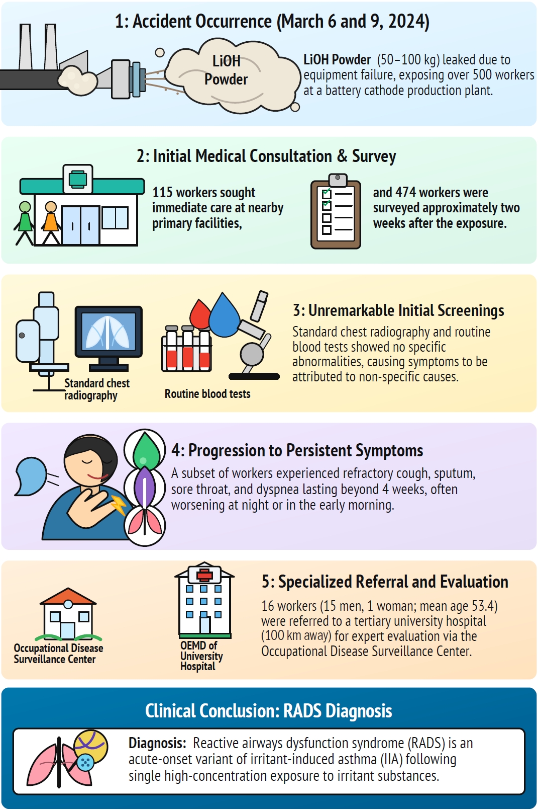
Selection pathway of the 16 workers from accident occurrence to evaluation at the Department of Occupational and Environmental Medicine (OEMD). The figure outlines the selection and diagnostic pathway following a major chemical exposure incident. In March 2024, a silicone connector in the powder supply line tore at a battery cathode material facility, releasing 50–100 kg of lithium hydroxide (LiOH) dust and exposing more than 500 workers. Many workers sought initial medical care, but routine evaluations—including chest X-rays and blood tests—showed no specific abnormalities, and their symptoms were often regarded as nonspecific. The figure then follows the subset of 16 workers whose respiratory symptoms persisted for over four weeks and who were referred to the OEMD at a tertiary hospital via the Occupational Disease Surveillance Center. The pathway ends with the clinical diagnosis of reactive airways dysfunction syndrome (RADS), highlighting the potential for long-term respiratory sequelae after high-concentration irritant exposure.
Table 1.
Demographic and clinical characteristics of 16 workers exposed to lithium hydroxide
| Case | Sex | Age (years) | Job | Visita | F/Ub | Smokingc | Comorbidities | WC | ||
|---|---|---|---|---|---|---|---|---|---|---|
| First | Last | No. | ||||||||
| A | M | 62 | General laborer | 2024 Apr 15 | 2026 Mar 25 | 33 | 23 | Never | HTN, DM | Approved |
| B | M | 58 | Pipe fitter | 2024 Jul 29 | 2024 Aug 30 | 3 | 1 | Never | DM, dyslipidemia | Not applied |
| C | M | 53 | Pipe fitter, team leader | 2024 Jul 29 | 2024 Aug 30 | 3 | 1 | Never | None | Not applied |
| D | M | 59 | General laborer | 2024 Aug 5 | 2026 Mar 25 | 19 | 20 | Never | HTN, DM | Approved |
| E | M | 51 | Site supervisor | 2024 Apr 5 | 2025 Sep 2 | 16 | 18 | Ex-smoker | HTN | Approved |
| F | M | 59 | Pipe fitter | 2024 Apr 8 | 2026 Feb 27 | 40 | 23 | Never | None | Approved |
| G | M | 54 | Pipe fitter | 2024 Apr 8 | 2026 Mar 18 | 44 | 23 | Never | None | Approved |
| H | M | 52 | Equipment engineer | 2024 May 21 | 2026 Mar 30 | 46 | 22 | Never | None | Approved |
| I | M | 48 | Scaffolder | 2024 May 8 | 2024 Dec 13 | 8 | 7 | Never | CRS | Approved |
| J | M | 55 | Scaffolder | 2024 Jul 23 | 2025 Mar 19 | 11 | 8 | Ex-smoker | None | Approved |
| K | M | 36 | Equipment worker | 2024 Jul 24 | 2024 Oct 10 | 3 | 3 | Current (15 py) | None | Not applied |
| L | M | 63 | Scaffolder | 2024 Jul 23 | 2024 Sep 4 | 4 | 2 | Current (6 py) | HTN | Not applied |
| M | M | 56 | Equipment worker | 2024 Jul 24 | 2024 Oct 10 | 3 | 3 | Current (34 py) | None | Not applied |
| N | F | 47 | Fire prevention | 2024 Jul 24 | 2024 Oct 10 | 3 | 3 | Never | RA | Not applied |
| O | M | 53 | General laborer | 2024 Aug 5 | 2024 Oct 10 | 3 | 2 | Ex-smoker | None | Not applied |
| P | M | 48 | Equipment worker | 2024 Aug 5 | 2024 Sep 27 | 3 | 2 | Never | None | Not applied |
The exposure incident occurred on March 6 and 9, 2024.
WC: workers’ compensation; HTN: hypertension; DM: diabetes mellitus; CRS: chronic rhinosinusitis; RA: rheumatoid arthritis.
aNumber of outpatient visits to the Department of Occupational and Environmental Medicine;
bFollow-up duration in months from the exposure incident to the last visit (as of March 31, 2026);
cSmoking history in pack-years (py).
Table 2.
Symptom profiles and clinical course of 16 workers exposed to lithium hydroxide
Table 3.
PFTs, methacholine challenge, FeNO, and HRCT findings in 16 workers exposed to lithium hydroxide
| Case | FEV1 (% pred)a | FEV1/FVC (%)b | FEF25–75 (% pred)c | BD FEV1 Δ%d | BD FEF25–75 Δ%d | TLC (% pred)e | DLco adj (% pred)f | Raw (cmH₂O/L/s)g | MCT PC20 (mg/mL)h | FeNO (ppb)i | PFT interpretation | HRCT findings | PNS |
|---|---|---|---|---|---|---|---|---|---|---|---|---|---|
| A | 59 | 71 | 50 | –9 | 19 | 71 | 45 | 1.57 | Positive (5.08, 23%) | – | Obstructive + restrictive; ↓↓DLco; BHR(+); ↓↓FEF25–75 | Subpleural nodules (LUL) | Y |
| B | 78 | 80 | 92 | –7 | 0 | 68 | 60 | 1.52 | Positive (1.36, 29%) | – | Mild obstruction; ↓↓DLco; ↓TLC; BHR(+); ↑Raw | ILD (probable UIP) | N |
| C | 69 | 72 | 52 | –5 | –8 | 85 | 73 | 2.55 | Positive (6.74, 21%) | – | Obstructive; ↓↓FEF25–75; BHR(+); ↓DLco; ↑↑Raw | Non-specific | N |
| D | 84 | 82 | 107 | 2 | 8 | 107 | 84 | 1.66 | N/P | – | Non-specific | Bronchopneumonia/ | N |
| E | 82 | 84 | 116 | –2 | 12 | 106 | 71 | 0.90 | Negative (>16, 18%) | 12 | Mild ↓DLco; MCT borderline | GGO (RUL); solid nodule (RUL) | Y |
| F | 93 | 77 | 96 | 10 | 24 | 89 | 77 | 1.18 | Negative (>16, 15%) | 24 | Mild ↓DLco; significant BD response | Subpleural nodules (RUL, LLL) | Y |
| G | 95 | 77 | 95 | 7 | 26 | 107 | 87 | 1.45 | Negative (>16, 13%) | 23 | Mild BD response in FEF25–75 | Non-specific | Y |
| H | 95 | 76 | 95 | 8 | 29 | 95 | 117 | 0.77 | Negative (>16, 11%) | – | Non-specific | Non-specific | N |
| I | 100 | 88 | 146 | 3 | 9 | 95 | 75 | 1.08 | N/P | – | Borderline DLco | Non-specific | N |
| J | 94 | 84 | 135 | –2 | 6 | 118 | 81 | 1.40 | Negative (>16, 6%) | – | Non-specific | Subpleural nodules | N |
| K | 78 | 81 | 73 | 3 | 17 | 92 | 67 | 1.40 | N/P | – | Mild ↓FEV1; ↓FEF25–75; ↓DLco | GGN 3 mm (RUL); r/o fibrosis, AAH | Y |
| L | 93 | 80 | 98 | 4 | 23 | 100 | 88 | 1.35 | Negative (>16, 19%) | – | Non-specific | Peripheral nodules (BULs) | Y |
| M | 95 | 83 | 120 | 1 | 1 | 115 | 80 | 1.57 | N/P | – | ↑Raw | GGO (RLL subpleural) | Y |
| N | 106 | 80 | 101 | 8 | 26 | 117 | 97 | 1.27 | N/P | – | Non-specific | Focal GGO (RUL) | Y |
| O | 83 | 79 | 95 | 7 | 38 | 112 | 94 | 1.25 | N/P | – | Significant FEF25–75 BD response | Subpleural nodule (RUL) | N |
| P | 90 | 83 | 103 | 1 | 11 | 85 | 91 | 0.86 | N/P | – | Non-specific | Subpleural nodule (RLL) | N |
FEV1, FEV1/FVC, and FEF25–75 are all values after bronchodilator administration.
PFT: pulmonary function test; HRCT: high-resolution computed tomography; BD: bronchodilator; PC20: provocative concentration causing a 20% decline in FEV1; PNS: paranasal sinus view X-ray; BHR: bronchial hyperresponsiveness; LUL: left upper lobe; ILD: interstitial lung disease; UIP: usual interstitial pneumonia; N/P: not performed; LLL: left lower lobe; GGO: ground-glass opacity; RUL: right upper lobe; GGN: ground-glass nodule; r/o: rule out; AAH: atypical adenomatous hyperplasia; BUL: bilateral upper lobes; RLL: right lower lobe.
aFEV1 (% pred): forced expiratory volume in 1 second; Reduced FEV1 may suggest airflow limitation; obstruction is defined by reduced FEV1/FVC;
bFEV1/FVC (%): ratio of FEV1 to forced vital capacity (FVC); <70% confirms obstructive pattern;
cFEF25–75 (% pred): forced expiratory flow at 25–75% of FVC; reflects small airway function; <65% suggests small airway disease;
dBD response (Δ%): change after bronchodilator; FEV1 ≥12% and ≥200 mL = significant; FEF25–75 ≥25% suggests small airway reversibility;
eTLC (% pred): total lung capacity; <80% indicates restriction;
fDLco adj (% pred): diffusing capacity adjusted for hemoglobin; <75% indicates impaired gas transfer;
gRaw: airway resistance; normal ≤1.5 cmH₂O/L/s; elevated values indicate increased airway resistance;
hMCT PC20: methacholine challenge; PC20 <4 = moderate–severe BHR, 4–16 = mild BHR, >16 = negative. The % of MCT PC20 is the rate of FEV1 reduction at the corresponding concentration;
iFeNO: fractional exhaled nitric oxide; <25 ppb normal, 25–50 intermediate, >50 high (eosinophilic inflammation).
Table 4.
RADS diagnostic criteria evaluation for 16 workers exposed to lithium hydroxide
| Case | (1) No prior resp. disease | (2) Single high-conc. exposure | (3) Onset ≤24 hours | (4) Symptoms ≥3 months | (5) Asthma-like symptoms | (6) Airflow limitation | (7) Bronchial hyperreactivity | (8) Other Dx excluded | RADS classificationa,b | Key features/remarks |
|---|---|---|---|---|---|---|---|---|---|---|
| A | O | O | O | O | O | O | O | O | Definite | Highest criteria fulfillment; MCT(+) with persistent symptoms over 23 months |
| 2nd leak incident | Within hours | 23+ mo | Cough/wheeze/dyspnea | Intermittent limitation | MCT(+) | '08: Definite | ||||
| PC20 5.08 | '19: Definite | |||||||||
| B | O | O | O | O | O | O | O | △ | Probable + ILD | MCT strongly positive (PC20 2 mg/mL); concomitant ILD (UIP) noted |
| Dust leak incident | Within hours | Persistent | Cough/dyspnea | ILD (UIP pattern) | MCT(+) | r/o ILD | '08: Probable + ILD | |||
| PC20 1.36 | '19: Definite + ILD | |||||||||
| C | O | △ | △ | △ | △ | O | O | O | Probable | MCT(+) confirming BHR; short observation period is a limitation |
| Repeated exposure | Gradual onset | ~1 mo | Cough/sputum | RML atelectasis | MCT(+) | '08: Probable | ||||
| observed | PC20 6.74 | '19: Borderline | ||||||||
| D | O | O | O | O | O | △ | – | O | Probable | Most criteria met (C1–C5, C8); MCT not performed; C6 under follow-up |
| Dust exposure | Within hours | 20+ mo | Cough/dyspnea/wheeze | Under follow-up | Not performed | '08: Probable | ||||
| '19: Unconfirmed | ||||||||||
| E | O | O | O | O | O | △ | △ | O | Probable | MCT discontinued due to FEV1 decline during testing, suggestive of BHR |
| Dust leak incident | Within hours | 18+ mo | Cough/dyspnea | FEV1 63%→ normalized | MCT borderline (PC20 16) | '08: Probable | ||||
| '19: Not met | ||||||||||
| F | O | O | O | O | O | △ | – | O | Possible | Symptom recurrence upon irritant re-exposure consistent with airway dysfunction |
| Dust leak incident | Within hours | 23+ mo | Cough/dyspnea/voice change | FEV1/FVC 76% (borderline) | MCT(–) | '08: Possible | ||||
| FeNO 24 | '19: Not met | |||||||||
| G | O | O | O | O | O | – | – | O | Possible | Persistent symptom pattern consistent with irritant-induced airway dysfunction |
| Dust leak incident | Within hours | 23+ mo | Cough/dyspnea/voice change | Under follow-up | MCT(–) (initial) | '08: Possible | ||||
| '19: Not met | ||||||||||
| H | O | O | O | O | O | – | – | O | Possible | Symptom relapse upon medication cessation suggests persistent airway hyperreactivity |
| 2nd leak incident | Within hours | 22+ mo | Cough/dyspnea/pharyngitis | Normal | MCT(–) | '08: Possible | ||||
| FeNO 17 | '19: Not met | |||||||||
| I | O | O | O | O | O | – | – | △ | Possible | Symptom exacerbation after repeated exposure; h/o CRS is confounding factor |
| 3rd repeated exposure | Within hours | 7+ mo | Cough/pharyngitis | Normal | Not performed | r/o CRS | '08: Possible | |||
| '19: Unconfirmed | ||||||||||
| J | O | O | O | O | O | – | – | O | Possible | Reduced exercise tolerance (dyspnea on 2 flights of stairs) |
| Dust leak incident | Within hours | 8+ mo | Cough/dyspnea/chest pain | Normal | MCT(–) | '08: Possible | ||||
| '19: Not met | ||||||||||
| K | O | △ | △ | O | O | O | – | △ | Possible | Mild obstructive defect on PFT; smoking history requires differentiation |
| Repeated exposure | Gradual onset | Persistent | Cough/dyspnea | Mild obstructive | Not performed | Smoking | '08: Possible | |||
| 15 py | '19: Unconfirmed | |||||||||
| L | O | △ | △ | X | △ | – | – | △ | Unlikely | Pulmonary embolism as complicating comorbidity; alternative etiology |
| Αcute + chronic combined | Gradual (after 1 month) | ~2 mo | Mainly cough/sputum | Normal | MCT(–) | PE | '08: Unlikely | |||
| occurred | '19: Not met | |||||||||
| M | O | △ | △ | O | O | – | – | △ | Unlikely | Heavy smoker (34 py); COPD must be excluded |
| Repeated exposure | Gradual onset | Persistent | Cough/dyspnea | Normal | Not performed | Smoking | '08: Unlikely | |||
| 34 py | '19: Unconfirmed | |||||||||
| N | O | △ | △ | O | △ | △ | – | △ | Unlikely | Predominantly upper airway/nasal symptoms; lower respiratory minimal |
| Repeated exposure | Gradual onset | Persistent | Mainly chest tightness | Minimal obstructive | Not performed | r/o RA | '08: Unlikely | |||
| '19: Unconfirmed | ||||||||||
| O | O | O | O | △ | △ | – | – | O | Unlikely | Mild symptoms with rapid resolution; returned to work |
| Direct dust contact | Within hours | ~2 mo | Mild cough/sputum | Normal | Not performed | '08: Unlikely | ||||
| '19: Unconfirmed | ||||||||||
| P | O | O | O | △ | X | – | – | O | Unlikely | Predominantly dermatological/neurological manifestations |
| 3 Exposures | Within hours | ~2 mo | Minimal respiratory | Normal | Not performed | '08: Unlikely | ||||
| '19: Unconfirmed |
Diagnostic frameworks:
1. Original Brooks Criteria (1985): Eight criteria (1–8) as listed above. All eight should be fulfilled for a definite diagnosis of RADS. Reference: Brooks et al. Chest 1985;88(3):376-84.3
2. ACCP Streamlined Criteria (2008): Six criteria (1) Absence of preceding respiratory illness/asthma; (2) Onset after single high-concentration exposure; (3) Onset of symptoms within 24 hours; (4) Positive test for bronchial hyperreactivity; (5) Airflow obstruction may or may not be present; (6) Exclusion of other disorders. Removed the standalone requirement for symptom persistence ≥3 months and relaxed the airflow obstruction requirement. Reference: Tarlo et al. Chest 2008;134(3 Suppl):1S-41S.4
3. Brooks Refined Criteria (2019): Five criteria (1) Absence of preceding disease; (2) Single high-concentration exposure; (3) Very high exposure (within 24 hours); (4) Symptom onset minutes to hours (<24 hours); (5) Positive PC20 <8 mg/mL. Emphasizes PC20 threshold for increased diagnostic precision. Reference: Brooks SM. Am J Biomed Sci Res 2019;6(3):205-8.9
O: criterion met; △: partially met/borderline; X: criterion not met; –: negative or normal finding.
resp.: respiratory; conc.: concentration; Dx: diagnosis; RADS: reactive airways dysfunction syndrome; MCT: methacholine challenge test; PC20: provocative concentration causing a 20% decline in FEV1; ILD: interstitial lung disease; UIP: usual interstitial pneumonia; RML: right middle lobe; BHR: bronchial hyperresponsiveness; FEV1: forced expiratory volume in 1 second; FVC: forced vital capacity; h/o: history of; CRS: chronic rhinosinusitis; PFT: pulmonary function test; PE: pulmonary embolism; COPD: chronic obstructive pulmonary disease; r/o: rule out; RA: rheumatoid arthritis.
aClassification row 1 (bold): based on Brooks 1985 original eight criteria (1–8). Row 2: '08 = American College of Chest Physicians (ACCP) 2008; '19 = Brooks 2019;
bDefinite = all/most criteria met with positive MCT; Probable = most criteria met, MCT borderline or not performed with strong clinical evidence; Possible = clinical pattern consistent but MCT negative or not performed; Unlikely = minimal respiratory symptoms or rapid resolution; Unconfirmed = MCT not performed, cannot be classified under the 2019 framework; Not met = MCT negative or PC20 ≥8 mg/mL; Insufficient data = workup incomplete.
- 1. Lee CG, Park SH, Kang JW, Hwang SW, Kim HN, Ko HK. Acute health effects of accidental exposure to lithium hydroxide at a battery material production plant. Ann Occup Environ Med 2026;38:e6.ArticlePubMedPMCPDF
- 2. Lee HY, Koh DH, Choi J, Won YL. Occupational skin diseases among cathode material workers in the Korean lithium-ion battery industry: a descriptive case series. Ann Occup Environ Med 2025;37:e29.ArticlePubMedPMCPDF
- 3. Brooks SM, Weiss MA, Bernstein IL. Reactive airways dysfunction syndrome (RADS): persistent asthma syndrome after high level irritant exposures. Chest 1985;88(3):376–84.ArticlePubMed
- 4. Tarlo SM, Balmes J, Balkissoon R, Beach J, Beckett W, Bernstein D, et al. Diagnosis and management of work-related asthma: American College of Chest Physicians Consensus Statement. Chest 2008;134(3 Suppl):1S–41S.ArticlePubMed
- 5. Shakeri MS, Dick FD, Ayres JG. Which agents cause reactive airways dysfunction syndrome (RADS)?: a systematic review. Occup Med (Lond) 2008;58(3):205–11.ArticlePubMed
- 6. Walters GI, Huntley CC. Updated review of reported cases of reactive airways dysfunction syndrome. Occup Med (Lond) 2020;70(7):490–5.ArticlePubMedPDF
- 7. Kipen HM, Blume R, Hutt D. Asthma experience in an occupational and environmental medicine clinic: low-dose reactive airways dysfunction syndrome. J Occup Med 1994;36(10):1133–7.ArticlePubMed
- 8. Brooks SM, Hammad Y, Richards I, Giovinco-Barbas J, Jenkins K. The spectrum of irritant-induced asthma: sudden and not-so-sudden onset and the role of allergy. Chest 1998;113(1):42–9.ArticlePubMed
- 9. Brooks SM. Concise details of RADS. Am J Biomed Sci Res 2019;6(3):205–8.Article
- 10. Vandenplas O, Wiszniewska M, Raulf M, de Blay F, Gerth van Wijk R, Moscato G, et al. EAACI position paper: irritant-induced asthma. Allergy 2014;69(9):1141–53.ArticlePubMed
- 11. Blanc PD, Annesi-Maesano I, Balmes JR, Cummings KJ, Fishwick D, Miedinger D, et al. The occupational burden of nonmalignant respiratory diseases. An official American Thoracic Society and European Respiratory Society statement. Am J Respir Crit Care Med 2019;199(11):1312–34.ArticlePubMedPMCPDF
- 12. Barber CM, Cullinan P, Feary J, Fishwick D, Hoyle J, Mainman H, et al. British Thoracic Society clinical statement on occupational asthma. Thorax 2022;77(5):433–42.ArticlePubMed
- 13. Lemiere C, Vandenplas O. Asthma in the workplace. In: Broaddus VC, Ernst JD, King TE Jr, editors. Murray and Nadel's Textbook of Respiratory Medicine. Philadelphia, PA: Elsevier; 2022, 1395–408.
- 14. Global Initiative for Asthma. 2025 Global strategy for asthma management and prevention. https://ginasthma.org/wp-content/uploads/2025/11/GINA-2025-Update-25_11_08-WMS.pdf. Updated 2025. Accessed February 28, 2026.
- 15. Lemiere C, Lavoie G, Doyen V, Vandenplas O. Irritant-Induced asthma. J Allergy Clin Immunol Pract 2022;10(11):2799–806.ArticlePubMed
- 16. Ronsmans S, Le Moual N, Dumas O. Update on irritant-induced occupational asthma. Curr Opin Allergy Clin Immunol 2023;23(2):63–9.ArticlePubMed
- 17. Bedoya Jaramillo JE, Vergara Trujillo RA, Alarcon Plaza JT, Vanegas Wilches AC, Forero Vasquez BN, Osorio Reyes WR. The clinical significance and translational implications of subclinical interstitial lung abnormalities in asymptomatic adults: a narrative review. Cureus 2025;17(9):e92255.ArticlePubMedPMC
- 18. Pye HO, Nenes A, Alexander B, Ault AP, Barth MC, Clegg SL, et al. The acidity of atmospheric particles and clouds. Atmos Chem Phys 2020;20(8):4809–88.ArticlePubMedPMC
- 19. Huynh HN, McNeill VF. Heterogeneous chemistry of CaCO3 aerosols with HNO3 and HCl. J Phys Chem A 2020;124(19):3886–95.ArticlePubMed
- 20. van Berlo D, Haberzettl P, Gerloff K, Li H, Scherbart AM, Albrecht C, et al. Investigation of the cytotoxic and proinflammatory effects of cement dusts in rat alveolar macrophages. Chem Res Toxicol 2009;22(9):1548–58.ArticlePubMed
- 21. Ogunbileje JO, Nawgiri RS, Anetor JI, Akinosun OM, Farombi EO, Okorodudu AO. Particles internalization, oxidative stress, apoptosis and pro-inflammatory cytokines in alveolar macrophages exposed to cement dust. Environ Toxicol Pharmacol 2014;37(3):1060–70.ArticlePubMed
- 22. Gharpure A, Heim JW 2nd, Vander Wal RL. Characterization and hazard identification of respirable cement and concrete dust from construction activities. Int J Environ Res Public Health 2021;18(19):10126.ArticlePubMedPMC
- 23. Gorguner M, Akgun M. Acute inhalation injury. Eurasian J Med 2010;42(1):28–35.Article
- 24. Shahpoury P, Zhang ZW, Arangio A, Celo V, Dabek-Zlotorzynska E, Harner T, et al. The influence of chemical composition, aerosol acidity, and metal dissolution on the oxidative potential of fine particulate matter and redox potential of the lung lining fluid. Environ Int 2021;148:106343.ArticlePubMedPMC
- 25. Widdicombe JH. Volume of airway surface liquid in health and disease. Am J Respir Crit Care Med 2002;165(11):1566.ArticlePDF
- 26. National Center for Biotechnology Information. Lithium hydroxide (PubChem). https://pubchem.ncbi.nlm.nih.gov/compound/3939. Updated 2004. Accessed April 1, 2026.
- 27. Shcherbakov VN, Lukashov YY, Lukashov YM. Electrolytical properties of solutions of lithium hydroxide at high temperatures and pressures. Therm Eng 2013;60(4):280–4.ArticlePDF
- 28. Zajac M, Dreano E, Edwards A, Planelles G, Sermet-Gaudelus I. Airway surface liquid pH regulation in airway epithelium: current understandings and gaps in knowledge. Int J Mol Sci 2021;22(7):3384.ArticlePubMedPMC
- 29. Morice AH, Millqvist E, Bieksiene K, Birring SS, Dicpinigaitis P, Domingo Ribas C, et al. ERS guidelines on the diagnosis and treatment of chronic cough in adults and children. Eur Respir J 2020;55(1):1901136.ArticlePubMedPMC
- 30. Lechien JR, Akst LM, Hamdan AL, Schindler A, Karkos PD, Barillari MR, et al. Evaluation and management of laryngopharyngeal reflux disease: state of the art review. Otolaryngol Head Neck Surg 2019;160(5):762–82.ArticlePubMedPDF
- 31. Skogstad M, Skorstad M, Lie A, Conradi HS, Heir T, Weisaeth L. Work-related post-traumatic stress disorder. Occup Med (Lond) 2013;63(3):175–82.ArticlePubMed
- 32. Bisson JI, Cosgrove S, Lewis C, Robert NP. Post-traumatic stress disorder. BMJ 2015;351:h6161.ArticlePubMed
REFERENCES
Figure & Data
REFERENCES
Citations
Citations to this article as recorded by 
 Cite
Cite XML Download
Reactive airways dysfunction syndrome following acute lithium hydroxide dust exposure at a battery material production plant: a case series of 16 workers
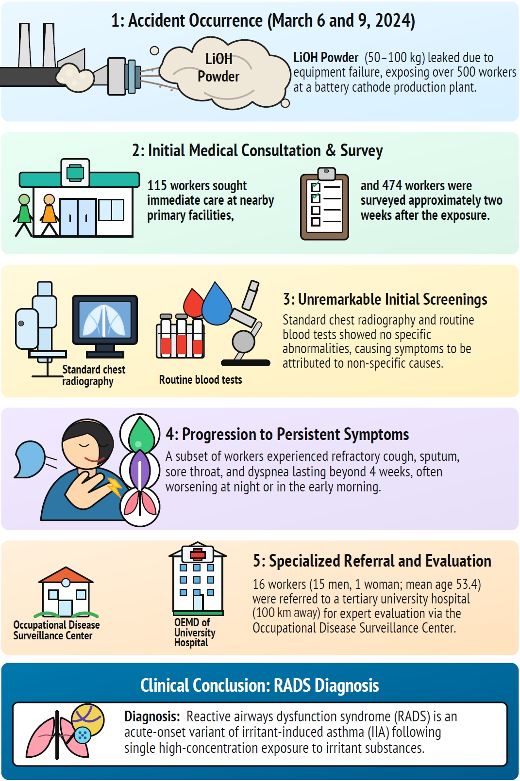
Fig. 1. Selection pathway of the 16 workers from accident occurrence to evaluation at the Department of Occupational and Environmental Medicine (OEMD). The figure outlines the selection and diagnostic pathway following a major chemical exposure incident. In March 2024, a silicone connector in the powder supply line tore at a battery cathode material facility, releasing 50–100 kg of lithium hydroxide (LiOH) dust and exposing more than 500 workers. Many workers sought initial medical care, but routine evaluations—including chest X-rays and blood tests—showed no specific abnormalities, and their symptoms were often regarded as nonspecific. The figure then follows the subset of 16 workers whose respiratory symptoms persisted for over four weeks and who were referred to the OEMD at a tertiary hospital via the Occupational Disease Surveillance Center. The pathway ends with the clinical diagnosis of reactive airways dysfunction syndrome (RADS), highlighting the potential for long-term respiratory sequelae after high-concentration irritant exposure.
Fig. 1.
Reactive airways dysfunction syndrome following acute lithium hydroxide dust exposure at a battery material production plant: a case series of 16 workers
| Case | Sex | Age (years) | Job | Visit |
F/U |
Smoking |
Comorbidities | WC | ||
|---|---|---|---|---|---|---|---|---|---|---|
| First | Last | No. | ||||||||
| A | M | 62 | General laborer | 2024 Apr 15 | 2026 Mar 25 | 33 | 23 | Never | HTN, DM | Approved |
| B | M | 58 | Pipe fitter | 2024 Jul 29 | 2024 Aug 30 | 3 | 1 | Never | DM, dyslipidemia | Not applied |
| C | M | 53 | Pipe fitter, team leader | 2024 Jul 29 | 2024 Aug 30 | 3 | 1 | Never | None | Not applied |
| D | M | 59 | General laborer | 2024 Aug 5 | 2026 Mar 25 | 19 | 20 | Never | HTN, DM | Approved |
| E | M | 51 | Site supervisor | 2024 Apr 5 | 2025 Sep 2 | 16 | 18 | Ex-smoker | HTN | Approved |
| F | M | 59 | Pipe fitter | 2024 Apr 8 | 2026 Feb 27 | 40 | 23 | Never | None | Approved |
| G | M | 54 | Pipe fitter | 2024 Apr 8 | 2026 Mar 18 | 44 | 23 | Never | None | Approved |
| H | M | 52 | Equipment engineer | 2024 May 21 | 2026 Mar 30 | 46 | 22 | Never | None | Approved |
| I | M | 48 | Scaffolder | 2024 May 8 | 2024 Dec 13 | 8 | 7 | Never | CRS | Approved |
| J | M | 55 | Scaffolder | 2024 Jul 23 | 2025 Mar 19 | 11 | 8 | Ex-smoker | None | Approved |
| K | M | 36 | Equipment worker | 2024 Jul 24 | 2024 Oct 10 | 3 | 3 | Current (15 py) | None | Not applied |
| L | M | 63 | Scaffolder | 2024 Jul 23 | 2024 Sep 4 | 4 | 2 | Current (6 py) | HTN | Not applied |
| M | M | 56 | Equipment worker | 2024 Jul 24 | 2024 Oct 10 | 3 | 3 | Current (34 py) | None | Not applied |
| N | F | 47 | Fire prevention | 2024 Jul 24 | 2024 Oct 10 | 3 | 3 | Never | RA | Not applied |
| O | M | 53 | General laborer | 2024 Aug 5 | 2024 Oct 10 | 3 | 2 | Ex-smoker | None | Not applied |
| P | M | 48 | Equipment worker | 2024 Aug 5 | 2024 Sep 27 | 3 | 2 | Never | None | Not applied |
| Case | Acute (≤24 hours) | Respiratory | Upper airway | Skin | Ocular | Systemic | Worsening | Course |
|---|---|---|---|---|---|---|---|---|
| A | Facial/cervical erythema, mucosal irritation | Cough (+), sputum (+), dyspnea (+), wheezing (+→improved) | Pharyngodynia, foreign body sensation; abnormal laryngoscopic findings | Facial/cervical erythema | None | Malaise (+), intermittent lower extremity edema | Yes (nocturnal) | Persistent (~2 years); fluctuating course with periodic exacerbation |
| B | Myalgia, exanthem, cough, sputum | Dry cough (+), sputum (+) | Pharyngolaryngeal foreign body sensation (NRS 3) | Persistent cervical/scalp/dorsal dermatitis | None | Severe malaise (+++), orthostatic vertigo | No | Partial resolution (residual symptoms) |
| C | Epistaxis (q 3–4 days), progressive nasal obstruction, increased sputum | Cough (+), sputum (+) (mild → controlled) | Pharyngolaryngeal foreign body sensation (NRS 3) | Pruritus/dermatitis | None | Severe malaise (+++), vertigo (“floating sensation”) | No | Partial resolution (pharmacotherapy adjustment ongoing) |
| D | Cough, sputum, pharyngodynia, dyspnea | Cough (++), sputum (++), dyspnea (++); gradual improvement | Pharyngodynia/foreign body sensation (+) | None | None | Malaise (+) | Yes (nocturnal/early AM) | Gradual resolution with persistent residual symptoms (~2 years) |
| E | Facial/truncal erythema, olfactory irritation | Cough (+), sputum (+), dyspnea (grade 1→worsened); early morning predominance | Pharyngodynia (NRS 5), persistent foreign body sensation | Erythematous papules/plaques (trunk, extremities) → residual scarring | None | Malaise (+), fatigue (+) | Yes (early AM) | Partial remission; chronic cough (>18 months duration) |
| F | Pharyngolaryngeal burning, cough, sputum, dysphonia | Cough (+), sputum (+), intermittent dyspnea (chest constriction) | Pharyngolaryngeal burning/foreign body sensation (NRS 4), dysphonia, dysphagia | Facial pruritus, periorbital edema | None | Malaise (+) | Yes (nocturnal awakening) | Persistent (~2 years); exacerbation upon irritant re-exposure |
| G | Cough, sputum, pharyngodynia, dysphonia | Cough (+), sputum (+), dyspnea (++); gradual improvement | Pharyngodynia/foreign body sensation (+), persistent dysphonia | None | None | Malaise (+→improved) | Yes (nocturnal) | Gradual resolution with persistent residual symptoms (~2 years) |
| H | Pharyngodynia (NRS 7–8), cough, sputum, intermittent dyspnea | Cough (+), sputum (+), dyspnea (+); gradual improvement | Pharyngodynia (NRS 7→3), foreign body sensation, dysphonia | None | None | Generalized fatigue (+), insomnia (+) | Yes (nocturnal) | Gradual resolution with persistent residual symptoms (~2 years) |
| I | Pharyngodynia (NRS 7), cough, sputum, febrile episode (exacerbated after 3rd exposure) | Cough (+++), sputum (+) | Pharyngodynia (NRS 7), dysphonia (+) | None | None | Vertigo (+), insomnia (+) | Not assessed | Persistent; transient improvement after HBO → re-exacerbation upon return to work |
| J | Pharyngeal globus sensation and pain | Cough (+), sputum (+), exertional dyspnea (onset climbing 2 flights) | Pharyngolaryngeal swelling (NRS 3→7→4), xerostomia, dysphonia | None | None | Malaise (+), myasthenia (+), depressed affect | Yes (AM/nocturnal) | Partial resolution with residual symptoms (AM predominance) |
| K | Epistaxis, insomnia, dyspnea, generalized fatigue | Cough (+), sputum (+), dyspnea (+++) | Pharyngolaryngeal foreign body sensation (NRS 3) | None | None | Severe malaise (+++), insomnia | No | Partial resolution (residual symptoms; returned to work) |
| L | Mild acute symptoms; progressive residual cough at 1 month post-exposure | Cough (+), hemoptysis-tinged sputum → PE diagnosed | Persistent oropharyngeal/pharyngolaryngeal discomfort | None | None | None | Yes (early AM) | Partial resolution (residual expectoration and nasal congestion) |
| M | Epistaxis, nasal mucosal hypertrophy, desquamation, generalized fatigue | Cough (+), sputum (+), intermittent dyspnea (early morning onset) | Pharyngolaryngeal pain (NRS 3)/foreign body sensation | Scalp epidermal desquamation (initial phase) | Visual blurring | Severe generalized fatigue (+++), vertigo (+) | Yes (early AM) | Partial resolution (residual symptoms) |
| N | Recurrent epistaxis, nasal mucosal hypertrophy | Mild cough, intermittent chest tightness | None (predominantly nasal symptoms) | Lower extremity/forearm contact dermatitis (late-onset) | None | Malaise (+) | No | Partial resolution (recurrent nasal mucosal inflammation) |
| O | Ocular foreign body sensation, pharyngolaryngeal pain | Cough (+), sputum (+) (mild) | Pharyngolaryngeal foreign body sensation (initial → resolved) | None | None | Malaise (+), pre-syncopal episodes (mild) | No | Resolved; returned to work (under surveillance) |
| P | Cephalalgia (persisting 3 weeks), tonsillitis, severe facial paresthesia | None (minimal respiratory symptoms) | None | Scalp seborrheic dermatitis (onset May–June) | None | Persistent fatigue, positional vertigo | No | Near-complete resolution (residual cephalalgia/vertigo) |
| Case | FEV1 (% pred) |
FEV1/FVC (%) |
FEF25–75 (% pred) |
BD FEV1 Δ% |
BD FEF25–75 Δ% |
TLC (% pred) |
DLco adj (% pred) |
Raw (cmH₂O/L/s) |
MCT PC20 (mg/mL) |
FeNO (ppb) |
PFT interpretation | HRCT findings | PNS |
|---|---|---|---|---|---|---|---|---|---|---|---|---|---|
| A | 59 | 71 | 50 | –9 | 19 | 71 | 45 | 1.57 | Positive (5.08, 23%) | – | Obstructive + restrictive; ↓↓DLco; BHR(+); ↓↓FEF25–75 | Subpleural nodules (LUL) | Y |
| B | 78 | 80 | 92 | –7 | 0 | 68 | 60 | 1.52 | Positive (1.36, 29%) | – | Mild obstruction; ↓↓DLco; ↓TLC; BHR(+); ↑Raw | ILD (probable UIP) | N |
| C | 69 | 72 | 52 | –5 | –8 | 85 | 73 | 2.55 | Positive (6.74, 21%) | – | Obstructive; ↓↓FEF25–75; BHR(+); ↓DLco; ↑↑Raw | Non-specific | N |
| D | 84 | 82 | 107 | 2 | 8 | 107 | 84 | 1.66 | N/P | – | Non-specific | Bronchopneumonia/ | N |
| E | 82 | 84 | 116 | –2 | 12 | 106 | 71 | 0.90 | Negative (>16, 18%) | 12 | Mild ↓DLco; MCT borderline | GGO (RUL); solid nodule (RUL) | Y |
| F | 93 | 77 | 96 | 10 | 24 | 89 | 77 | 1.18 | Negative (>16, 15%) | 24 | Mild ↓DLco; significant BD response | Subpleural nodules (RUL, LLL) | Y |
| G | 95 | 77 | 95 | 7 | 26 | 107 | 87 | 1.45 | Negative (>16, 13%) | 23 | Mild BD response in FEF25–75 | Non-specific | Y |
| H | 95 | 76 | 95 | 8 | 29 | 95 | 117 | 0.77 | Negative (>16, 11%) | – | Non-specific | Non-specific | N |
| I | 100 | 88 | 146 | 3 | 9 | 95 | 75 | 1.08 | N/P | – | Borderline DLco | Non-specific | N |
| J | 94 | 84 | 135 | –2 | 6 | 118 | 81 | 1.40 | Negative (>16, 6%) | – | Non-specific | Subpleural nodules | N |
| K | 78 | 81 | 73 | 3 | 17 | 92 | 67 | 1.40 | N/P | – | Mild ↓FEV1; ↓FEF25–75; ↓DLco | GGN 3 mm (RUL); r/o fibrosis, AAH | Y |
| L | 93 | 80 | 98 | 4 | 23 | 100 | 88 | 1.35 | Negative (>16, 19%) | – | Non-specific | Peripheral nodules (BULs) | Y |
| M | 95 | 83 | 120 | 1 | 1 | 115 | 80 | 1.57 | N/P | – | ↑Raw | GGO (RLL subpleural) | Y |
| N | 106 | 80 | 101 | 8 | 26 | 117 | 97 | 1.27 | N/P | – | Non-specific | Focal GGO (RUL) | Y |
| O | 83 | 79 | 95 | 7 | 38 | 112 | 94 | 1.25 | N/P | – | Significant FEF25–75 BD response | Subpleural nodule (RUL) | N |
| P | 90 | 83 | 103 | 1 | 11 | 85 | 91 | 0.86 | N/P | – | Non-specific | Subpleural nodule (RLL) | N |
| Case | (1) No prior resp. disease | (2) Single high-conc. exposure | (3) Onset ≤24 hours | (4) Symptoms ≥3 months | (5) Asthma-like symptoms | (6) Airflow limitation | (7) Bronchial hyperreactivity | (8) Other Dx excluded | RADS classification |
Key features/remarks |
|---|---|---|---|---|---|---|---|---|---|---|
| A | O | O | O | O | O | O | O | O | Definite | Highest criteria fulfillment; MCT(+) with persistent symptoms over 23 months |
| 2nd leak incident | Within hours | 23+ mo | Cough/wheeze/dyspnea | Intermittent limitation | MCT(+) | '08: Definite | ||||
| PC20 5.08 | '19: Definite | |||||||||
| B | O | O | O | O | O | O | O | △ | Probable + ILD | MCT strongly positive (PC20 2 mg/mL); concomitant ILD (UIP) noted |
| Dust leak incident | Within hours | Persistent | Cough/dyspnea | ILD (UIP pattern) | MCT(+) | r/o ILD | '08: Probable + ILD | |||
| PC20 1.36 | '19: Definite + ILD | |||||||||
| C | O | △ | △ | △ | △ | O | O | O | Probable | MCT(+) confirming BHR; short observation period is a limitation |
| Repeated exposure | Gradual onset | ~1 mo | Cough/sputum | RML atelectasis | MCT(+) | '08: Probable | ||||
| observed | PC20 6.74 | '19: Borderline | ||||||||
| D | O | O | O | O | O | △ | – | O | Probable | Most criteria met (C1–C5, C8); MCT not performed; C6 under follow-up |
| Dust exposure | Within hours | 20+ mo | Cough/dyspnea/wheeze | Under follow-up | Not performed | '08: Probable | ||||
| '19: Unconfirmed | ||||||||||
| E | O | O | O | O | O | △ | △ | O | Probable | MCT discontinued due to FEV1 decline during testing, suggestive of BHR |
| Dust leak incident | Within hours | 18+ mo | Cough/dyspnea | FEV1 63%→ normalized | MCT borderline (PC20 16) | '08: Probable | ||||
| '19: Not met | ||||||||||
| F | O | O | O | O | O | △ | – | O | Possible | Symptom recurrence upon irritant re-exposure consistent with airway dysfunction |
| Dust leak incident | Within hours | 23+ mo | Cough/dyspnea/voice change | FEV1/FVC 76% (borderline) | MCT(–) | '08: Possible | ||||
| FeNO 24 | '19: Not met | |||||||||
| G | O | O | O | O | O | – | – | O | Possible | Persistent symptom pattern consistent with irritant-induced airway dysfunction |
| Dust leak incident | Within hours | 23+ mo | Cough/dyspnea/voice change | Under follow-up | MCT(–) (initial) | '08: Possible | ||||
| '19: Not met | ||||||||||
| H | O | O | O | O | O | – | – | O | Possible | Symptom relapse upon medication cessation suggests persistent airway hyperreactivity |
| 2nd leak incident | Within hours | 22+ mo | Cough/dyspnea/pharyngitis | Normal | MCT(–) | '08: Possible | ||||
| FeNO 17 | '19: Not met | |||||||||
| I | O | O | O | O | O | – | – | △ | Possible | Symptom exacerbation after repeated exposure; h/o CRS is confounding factor |
| 3rd repeated exposure | Within hours | 7+ mo | Cough/pharyngitis | Normal | Not performed | r/o CRS | '08: Possible | |||
| '19: Unconfirmed | ||||||||||
| J | O | O | O | O | O | – | – | O | Possible | Reduced exercise tolerance (dyspnea on 2 flights of stairs) |
| Dust leak incident | Within hours | 8+ mo | Cough/dyspnea/chest pain | Normal | MCT(–) | '08: Possible | ||||
| '19: Not met | ||||||||||
| K | O | △ | △ | O | O | O | – | △ | Possible | Mild obstructive defect on PFT; smoking history requires differentiation |
| Repeated exposure | Gradual onset | Persistent | Cough/dyspnea | Mild obstructive | Not performed | Smoking | '08: Possible | |||
| 15 py | '19: Unconfirmed | |||||||||
| L | O | △ | △ | X | △ | – | – | △ | Unlikely | Pulmonary embolism as complicating comorbidity; alternative etiology |
| Αcute + chronic combined | Gradual (after 1 month) | ~2 mo | Mainly cough/sputum | Normal | MCT(–) | PE | '08: Unlikely | |||
| occurred | '19: Not met | |||||||||
| M | O | △ | △ | O | O | – | – | △ | Unlikely | Heavy smoker (34 py); COPD must be excluded |
| Repeated exposure | Gradual onset | Persistent | Cough/dyspnea | Normal | Not performed | Smoking | '08: Unlikely | |||
| 34 py | '19: Unconfirmed | |||||||||
| N | O | △ | △ | O | △ | △ | – | △ | Unlikely | Predominantly upper airway/nasal symptoms; lower respiratory minimal |
| Repeated exposure | Gradual onset | Persistent | Mainly chest tightness | Minimal obstructive | Not performed | r/o RA | '08: Unlikely | |||
| '19: Unconfirmed | ||||||||||
| O | O | O | O | △ | △ | – | – | O | Unlikely | Mild symptoms with rapid resolution; returned to work |
| Direct dust contact | Within hours | ~2 mo | Mild cough/sputum | Normal | Not performed | '08: Unlikely | ||||
| '19: Unconfirmed | ||||||||||
| P | O | O | O | △ | X | – | – | O | Unlikely | Predominantly dermatological/neurological manifestations |
| 3 Exposures | Within hours | ~2 mo | Minimal respiratory | Normal | Not performed | '08: Unlikely | ||||
| '19: Unconfirmed |
Table 1. Demographic and clinical characteristics of 16 workers exposed to lithium hydroxide
The exposure incident occurred on March 6 and 9, 2024. WC: workers’ compensation; HTN: hypertension; DM: diabetes mellitus; CRS: chronic rhinosinusitis; RA: rheumatoid arthritis. Number of outpatient visits to the Department of Occupational and Environmental Medicine; Follow-up duration in months from the exposure incident to the last visit (as of March 31, 2026); Smoking history in pack-years (py).
Table 2. Symptom profiles and clinical course of 16 workers exposed to lithium hydroxide
NRS: Numeric Rating Scale; PE: pulmonary embolism; HBO: hyperbaric oxygen therapy; AM: morning; (+): mild; (++): moderate; (+++): severe.
Table 3. PFTs, methacholine challenge, FeNO, and HRCT findings in 16 workers exposed to lithium hydroxide
FEV1, FEV1/FVC, and FEF25–75 are all values after bronchodilator administration. PFT: pulmonary function test; HRCT: high-resolution computed tomography; BD: bronchodilator; PC20: provocative concentration causing a 20% decline in FEV1; PNS: paranasal sinus view X-ray; BHR: bronchial hyperresponsiveness; LUL: left upper lobe; ILD: interstitial lung disease; UIP: usual interstitial pneumonia; N/P: not performed; LLL: left lower lobe; GGO: ground-glass opacity; RUL: right upper lobe; GGN: ground-glass nodule; r/o: rule out; AAH: atypical adenomatous hyperplasia; BUL: bilateral upper lobes; RLL: right lower lobe. FEV1 (% pred): forced expiratory volume in 1 second; Reduced FEV1 may suggest airflow limitation; obstruction is defined by reduced FEV1/FVC; FEV1/FVC (%): ratio of FEV1 to forced vital capacity (FVC); <70% confirms obstructive pattern; FEF25–75 (% pred): forced expiratory flow at 25–75% of FVC; reflects small airway function; <65% suggests small airway disease; BD response (Δ%): change after bronchodilator; FEV1 ≥12% and ≥200 mL = significant; FEF25–75 ≥25% suggests small airway reversibility; TLC (% pred): total lung capacity; <80% indicates restriction; DLco adj (% pred): diffusing capacity adjusted for hemoglobin; <75% indicates impaired gas transfer; Raw: airway resistance; normal ≤1.5 cmH₂O/L/s; elevated values indicate increased airway resistance; MCT PC20: methacholine challenge; PC20 <4 = moderate–severe BHR, 4–16 = mild BHR, >16 = negative. The % of MCT PC20 is the rate of FEV1 reduction at the corresponding concentration; FeNO: fractional exhaled nitric oxide; <25 ppb normal, 25–50 intermediate, >50 high (eosinophilic inflammation).
Table 4. RADS diagnostic criteria evaluation for 16 workers exposed to lithium hydroxide
Diagnostic frameworks: 1. Original Brooks Criteria (1985): Eight criteria (1–8) as listed above. All eight should be fulfilled for a definite diagnosis of RADS. Reference: Brooks et al. Chest 1985;88(3):376-84. 2. ACCP Streamlined Criteria (2008): Six criteria (1) Absence of preceding respiratory illness/asthma; (2) Onset after single high-concentration exposure; (3) Onset of symptoms within 24 hours; (4) Positive test for bronchial hyperreactivity; (5) Airflow obstruction may or may not be present; (6) Exclusion of other disorders. Removed the standalone requirement for symptom persistence ≥3 months and relaxed the airflow obstruction requirement. Reference: Tarlo et al. Chest 2008;134(3 Suppl):1S-41S. 3. Brooks Refined Criteria (2019): Five criteria (1) Absence of preceding disease; (2) Single high-concentration exposure; (3) Very high exposure (within 24 hours); (4) Symptom onset minutes to hours (<24 hours); (5) Positive PC20 <8 mg/mL. Emphasizes PC20 threshold for increased diagnostic precision. Reference: Brooks SM. Am J Biomed Sci Res 2019;6(3):205-8. O: criterion met; △: partially met/borderline; X: criterion not met; –: negative or normal finding. resp.: respiratory; conc.: concentration; Dx: diagnosis; RADS: reactive airways dysfunction syndrome; MCT: methacholine challenge test; PC20: provocative concentration causing a 20% decline in FEV1; ILD: interstitial lung disease; UIP: usual interstitial pneumonia; RML: right middle lobe; BHR: bronchial hyperresponsiveness; FEV1: forced expiratory volume in 1 second; FVC: forced vital capacity; h/o: history of; CRS: chronic rhinosinusitis; PFT: pulmonary function test; PE: pulmonary embolism; COPD: chronic obstructive pulmonary disease; r/o: rule out; RA: rheumatoid arthritis. Classification row 1 (bold): based on Brooks 1985 original eight criteria (1–8). Row 2: '08 = American College of Chest Physicians (ACCP) 2008; '19 = Brooks 2019; Definite = all/most criteria met with positive MCT; Probable = most criteria met, MCT borderline or not performed with strong clinical evidence; Possible = clinical pattern consistent but MCT negative or not performed; Unlikely = minimal respiratory symptoms or rapid resolution; Unconfirmed = MCT not performed, cannot be classified under the 2019 framework; Not met = MCT negative or PC20 ≥8 mg/mL; Insufficient data = workup incomplete.


