Factors related to femoral bowing among Korean female farmers: a cross-sectional study
Article information

Abstract
Background
Female farmers have a high prevalence of knee osteoarthritis (KOA) in South Korea. Femoral bowing has been reported to be related to KOA by increasing load on the mechanical axis. This study aimed to investigate factors related to femoral bowing in Korean female farmers.
Methods
We analyzed the legs of 264 female farmers registered with the Korea farmers' knee cohort of Jeonnam Center for Farmers' Safety and Health. A structured questionnaire was used to determine sociodemographic variables, agricultural career, cumulative squatting working time (CSWT), and cumulative heavy lifting working time. Femoral bone density was measured and Kellgren-Lawrence (KL) grades were obtained from the knee radiographs. Mechanical axis angle (MAA), femoral bowing angle (FBA), anatomical lateral distal femoral angle (aLDFA), anatomical medial proximal tibial angle (aMPTA), and condylar-plateau angle (CPA) were measured. We examined the relationship between the FBA and related factors by using multiple linear regression.
Results
The proportion of individuals with radiographic KOA (≥ KL grade 2) in this study was 37.9%. As KL grades increased, MAA, FBA, and CPA increased, whereas aLDFA and aMPTA decreased. FBA increased with age. Multiple linear regression analyses using FBA as a dependent variable showed relationship with higher age, lower height, higher BMI, lower bone mineral density, longer CSWT, and longer agricultural careers.
Conclusions
The results of this study suggest that external factors related to agricultural work in female farmers was associated with femoral bowing, in addition to internal factors such as age, bone density, height, and obesity.
BACKGROUND
Osteoarthritis is a chronic degenerative disorder that causes anatomic and physiologic derangement. Its global prevalence is increasing and is expected to increase continuously [1]. Osteoarthritis is a significant economic burden for individuals and is often a cause of ‘catastrophic health expenditure’ as defined by the World Health Organization [2]. Knee osteoarthritis (KOA) is significant, in comparison to arthritis affecting other joints, as it affects ambulation and social function [34]. Females have a higher prevalence of osteoarthritis than males [5]. This trend was seen in the fifth Korea National Health and Nutrition Examination Survey, which estimated that 26.7% of men and 47.3% of women have radiographic KOA. The rate of total knee arthroplasties has increased steadily from 2001 to 2010 in both sexes and the proportion of female patients has been steady at approximately 90% [6]. Female farmers in South Korea have a higher prevalence than females in other occupations [7]. The high prevalence is thought to be due to occupational factors, such as squatting, climbing, and lifting in addition to the biological vulnerability of women [89]. Various hypotheses have been suggested for the high prevalence of KOA in women [101112]. The malalignment of lower extremities as intrinsic factors of KOA is critical because it fails to distribute weight evenly [131415]. Varus deformity is the result of bone deformation with age and progressive KOA [151617]. The purpose of this study was to examine the epidemiological characteristics of lower limb alignment in female farmers and to determine whether squatting or physical burden might contribute to the change of lower limb alignment in female farmers. In particular, the coronal femoral bowing angle (FBA) has recently been noted as the major contributing factor to the mechanical axis angle (MAA) in females [1819202122]. Some researchers have hypothesized that the FBA is associated with the progression of KOA [2324]. Therefore, we analyzed the characteristics of femoral deformation in Korean female farmers. We also identified the external and internal factors associated with the FBA.
METHODS
Participants
This cross-sectional study enrolled farmers registered with the Korea farmers' knee cohort (KFKC). This cohort consists of farmers from 16 districts in Jeonnam province, South Korea. We analyzed 528 knees of 264 women from this cohort aged 40–79 years, between 2013 and 2015. The subjects who underwent arthroplasty were excluded from the screening process in the KFKC because the KFKC is a magnetic resonance imaging-based cohort.
Related variables
Researchers obtained information on sex and age, physical profiles such as height and body mass index (BMI), working time and length of farming career, using the structured questionnaire. They obtained the bone mineral density measures and knee radiographs (weight-bearing whole leg anteroposterior view) from the KFKC database. To measure how much time the farmers spent working in a specific position (squatting or lifting), we took the unit task exposure as the median value of each sub-periods exposure and then defined cumulative squatting working time (CSWT) and cumulative heavy lifting working time (CLWT) by the following formula: cumulative working time (hours) = working years for lifetime × working months per year × 4 weeks × working days per week × working hours per day. However, right-skewed distribution was found in the initial analysis of CSWT and CLWT, so we converted the value of these variables into quartiles (4Q) for regression analysis. The bone marrow density data in the KFKC database were the sets of values measured at the lumbar spine (L1 to L4) and femoral neck. However, for this study, we used the value of femur bone mineral density (BMD[f]).
Radiographic measurements
The Kellgren-Lawrence (KL) grade was used to determine the stage of KOA [25]. Two experienced physicians read the images taken with standardized weight-bearing for anteroposterior and Rosenburg view. We used an atlas from the Osteoarthritis Research Society International to increase the reliability of the readings [26], and 4 mm was applied as the cut-off point for joint space narrowing, based on the International Knee Documentation Committee [2527]. The MAA was defined as the angle between one line connecting the center of the femoral head with the middle of the knee (midpoint of the tibial spine) and another line connecting the middle of the surface of the talus with the middle of the knee [28]. The FBA was defined as an acute angle formed between the line drawn at the center of the femur below the level of the lesser trochanter passing through the center of the femur at a point 5 cm distal to the starting point and the line extending from the center of the femoral distal condyle through the center of the femur at a 5 cm proximal portion and a 5 cm further proximal point. The lateral FBA was expressed as a positive value [29]. The anatomical lateral distal femoral angle (aLDFA) was the angle between the anatomical axis of the femur and the tangent to the subchondral plate of the femoral condyle. The anatomical medial proximal tibial angle (aMPTA) was the angle between the tangent to the subchondral plate of the tibia and the anatomical axis of the tibia [30]. The measuring point for the anatomical shaft axis was 10 cm away from the joint surface. The condylar plateau angle (CPA) was the angle between the tangents to the subchondral plates of the femoral and tibial condyles (Fig. 1) [31].
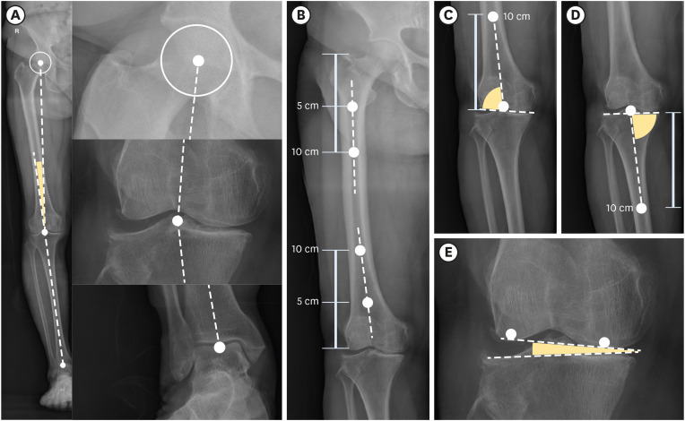
Measurement of MAA and lower limb angles. (A) MAA, (B) femoral bowing angle, (C) anatomical lateral distal femoral angle, (D) anatomical medial tibial angle, (E) condylar plateau angle.
MAA: mechanical axis angle.
Reliability of radiologic measurement
We calculated intraclass correlation coefficients (ICC) for interobserver reliability. The ICC of MAA was excellent (ICC 0.981) and the ICC of FBA and CPA was good (0.809 and 0.842 respectively). The ICCs of aLDFA and aMPTA, however, were moderate, at 0.561 and 0.695 respectively. The Kappa of the KL grade by 2 readers was moderate (kappa 0.581). The value measured by the more experienced reader was selected, and when the difference between the measured values was large, the re-measured value was used.
Statistical analysis
We performed 1-way analysis of variance and post hoc analysis by Scheffé's test to compare the mean values of the lower limb angles according to the KL grade group. The linear regression analysis with MAA as the dependent variable and FBA, aLDFA, CPA, and aMPTA as independent variables applied for estimating the contribution of lower limb angles for MAA. The relationship between the variables was obtained by Pearson's correlation analysis. To evaluate the association between the FBA and related factors, a linear regression analysis was performed with 2 models, to reduce the problem of multicollinearity. Model 1 included internal factors such as age, height, BMI, and BMD(f), and agricultural work-related external factors such as CSWT and CLWT. In model 2, CSWT, and CLWT were replaced by agricultural career All statistical analyses were performed using IBM SPSS version 21.0 (IBM Corp., Armonk, NY, USA). A p-value of < 0.05 was considered statistically significant.
Ethics statement
The present study protocol was reviewed and approved by the Institutional Review Board of Chosun University Hospital (approval No. 2013-12-006). Informed consent was submitted by all subjects when they were enrolled. All participants provided written informed consent for both the participation in the survey and the use of their data for research purposes.
RESULTS
General characteristics
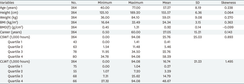
The average age of the study participants was 57.27 years (± 8.19). The average height was 155.57 (± 6.36) cm, and the average weight was 59.01 (± 9.08) kg. The average BMI and BMD(f) were 24.31 kg/m2 (± 3.15) and 0.92 g/cm2 (± 0.14), respectively. The average duration of agricultural career was 27.05 years (± 15.21). The average CSWT was 25.03 thousand hours, with a skewness of 0.893. The average CLWT was 21.23 thousand hours, with a skewness of 1.495 (Table 1).
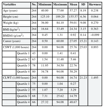
General characteristics
Comparison of the means of the lower limb angles by severity of ROA
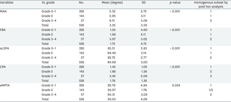
Table 2 shows the comparison of the mean values of the lower limb angles according to the KL grade. As KL grade increased, MAA, FBA, and CPA increased significantly, whereas aLDFA and aMPFA decreased significantly. In post hoc tests, MAA, FBA, and aLDFA were divided into KL grades 0–2 and 3–4. In contrast, CPA showed significant differences in KL grades 0–1, grade 2, and grades 3–4, respectively.
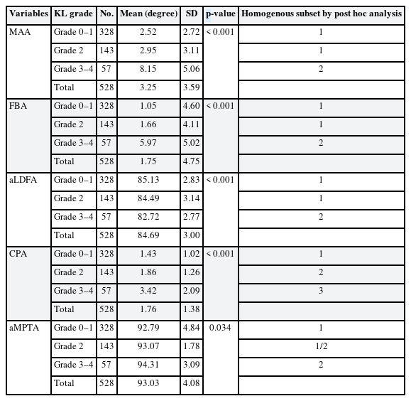
Comparison of means of lower limb angles according to KL grade group
The correlation between the lower limb angles
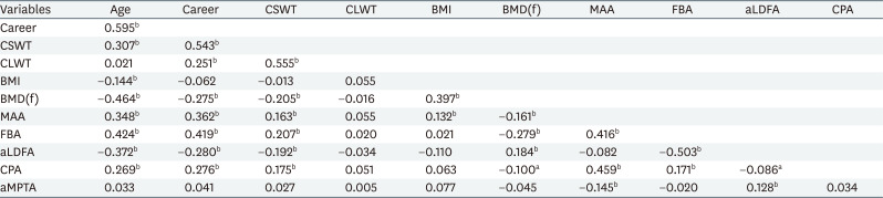
Table 3 shows the association between the variables through Pearson's correlation analysis. The variables highly correlated with age were the agricultural career (correlation coefficient [r] = 0.595), BMD(f) (r = 0.464), FBA (r = 0.424), MMA (r = 0.348), aLDFA (r = −0.372), CSWT (r = 0.307), and CPA (r = 0.269). Agricultural career was correlated with CSWT (r = 0.543) and CLWT (r = 0.251). CLWT and CSWT were also correlated (r = 0.555). MAA showed a significant positive correlation with FBA (r = 0.416) and CPA (r = 0.459), whereas aLDFA had no correlation and aMPTA had a significant negative correlation (r = −0.145). FBA showed a significant negative correlation with aLDFA (r = −0.503) and a significant positive correlation with CPA (r = 0.171) (Fig. 2).
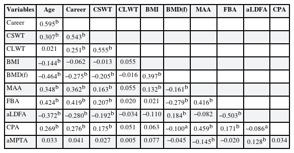
The correlation coefficient between factors
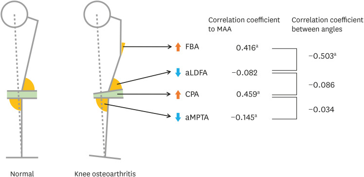
The change of lower limb angles from normal to knee osteoarthritis.
MAA: mechanical axis angle; FBA: femoral bowing angle; aLDFA: anatomical lateral distal femoral angle; CPA: condylar-plateau angle (joint space narrowing); aMPTA: anatomical medial proximal tibial angle.
ap < 0.01 by Pearson's correlation analysis.
Factors associated with the FBA
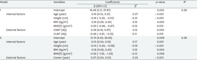
Table 4 shows the result of multiple linear regression analysis using FBA as a dependent variable. In model 1, FBA was positively correlated with increase in age, BMI, and CSWT. On the other hand, as the values of height, BMD(f), and CLWT (4Q) increased, FBA significantly decreased. In model 2, FBA was positively correlated with increase in age, BMI, and career. Likewise, as the height, BMD(f) and CLWT (4Q) increased, FBA significantly decreased. In model 1, the standardized coefficient beta (β*) was in order of age (β* = 0.27), height (β* = −0.21), and CSWT (β* = 0.13). However, in model 2, the standardized beta was in order of agricultural career (β* = 0.23), height (β* = −0.19), and age (β* = 0.17).
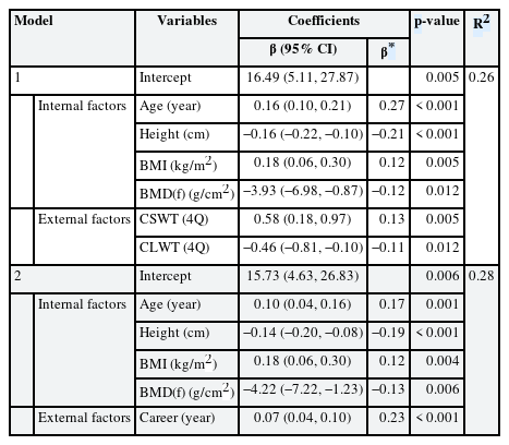
The related factors of femoral bowing angle by multiple linear regression
DISCUSSION
It is well known that the CPA is associated with KOA [32], and this has been confirmed in our study. A higher CPA indicates that the medial tibiofemoral joint space is reduced compared to the lateral side. Therefore, medial compartment osteoarthritis leads to an increase in the CPA. In our study, age and BMI were related to the CPA, as in previous studies [182324].
In our study, we identified a significant association between the FBA and internal factors such as age, height, BMI, and BMD(f). In particular, we found an association of the FBA with occupational external factors such as prolonged squatting posture or agricultural career in Korean female farmers after the adjustment for known confounders. These results suggest that femoral bowing observed in female farmers is related to agricultural work.
The results of our study show that the FBA increases with age. However, an additional analysis comparing both sexes showed that this phenomenon only occurred in female farmers. The association between the FBA and age was similar, although subjects were limited to only persons with radiologic KOA. Interestingly, in female farmers without radiologic KOA, the FBA increased with age (Supplementary Table 1). In interpreting these phenomena, Matsumoto et al.'s [23] almost unique population-based study has become an important comparison with ours. They conducted a cross-sectional study comparing limb alignment between normal volunteers and KOA patients who visited Kobe university in Japan. They also found that the femoral shaft bowed medially with age. However, the relationship between age and the FBA in normal people did not show a significant difference between males and females. The increase in the FBA was more prominent in females than in males in KOA patients [23]. This difference is presumed to be due to the different characteristics of the study subjects. Matsumoto et al.'s [23] subjects were recruited by voluntary participation from the community, but our research subjects were limited to agricultural workers.
Several studies have shown that the FBA of patients with KOA is larger than that of the normal control group [24293334353637]. These results have been reported mainly in hospital-based studies. Matsumoto et al. [23] noted the possibility that an increase in the FBA triggers or worsens KOA. They showed that the mean FBA among females with mild KOA was 1.4 ± 3.1, but the mean FBA among profound KOA patients was 5.9 ± 3.2 [23]. In addition, the FBA increased with the severity of KOA. Yau et al. [37] suggested that femoral bowing was common in a Chinese population with end-stage KOA.
Our study showed the higher the BMD and height, the lower the FBA. Shimosawa et al. [24] also showed similar results. Rickets and osteomalacia induced by poor dietary intake of vitamin D, present with bowing of the legs. In particular, vitamin D is related to the growth hormone/insulin-like growth factor 1 axis in childhood [38]. Therefore, vitamin D or nutritional deficiency in childhood is a shared factor with lower peak bone density and lower height. Although the relationship between age and FBA is not clear, it is assumed that factors such as malnutrition and menopause have been mediated. Our study did not investigate vitamin D deficiency, but there is sufficient evidence that vitamin D deficiency causes low BMD. Insufficient daily calcium and vitamin D intake is common in Korean postmenopausal women [39]. The softened bones of females with osteomalacia can lead to bowing, especially in the weight-bearing bones of the legs [40].
In addition to confirming this fact, our study identified which external factors cause FBA in vulnerable women. Our conclusion is that prolonged squatting increases FBA, and heavy lifting decreases FBA. The squatting position of Korean female farmers, called Asian squat, is a deep squatting posture with spreading between both knees and contact between the thighs and calves [41]. This posture applies force perpendicular to the proximal femur, whereas, heavy lifting in the standing position puts load along the axis of the long bones. In addition, heavy lifting is a resistance exercise that improves lower extremity muscles, and thus has a positive effect on bone density [42]. Shin et al. [20] showed the association between atypical femoral fractures with femoral bowing and loss of thigh muscle. These results support the conclusion that lifting maintains or strengthens muscles and can be a protective factor against femoral bowing.
Agricultural career was significantly related to the FBA even after adjusting age. In model 1, the standardized beta of age was the largest, at 0.27. In model 2, the standardized beta of age was reduced to 0.17 and the standardized beta of the agricultural career was the largest, at 0.23. This suggests that there may be external factors related to agricultural work that are not explained by CSWT and CLWT.
Interestingly, our study showed a clear negative correlation between the FBA and aLDFA (r = −0.503). FBA and aMPTA showed no correlation. As mentioned earlier, FBA has a positive correlation with the severity of KOA. According to a study by Matsumoto et al. [23], The severity of KOA and aLDFA have a positive correlation. This trend was also confirmed in our study. Although both FBA and aLDFA are related to the severity of radiologic KOA, it is unusual to have an inverse correlation between FBA and aLDFA. However, this result was also reproduced in the study by Lu et al. [33].
The femur is a long bone, while the femur condyle is a relatively wide and thick bone. Thus, when bone deformation is caused by external or internal factors, the femur's deformation will be greater than that of the condyle. We thus interpreted that as the FBA increased, aLDFA, the angle between the femur shaft and the femur condyle, also increased.
This study has the following limitations. First, since participants are female farmers living in a rural society, the results cannot be generalized to males, urban residents, and other occupational groups. Second, CLWT and CSWT were insufficient to verify reliability and validity. The CLWT and CSWT had a bimodal and right-skewed distribution, we, therefore, categorized the cumulative working times into 4Q. Third, survival bias is possible because people with knee arthroplasty were excluded from the study. Fourth, calcium or vitamin D intake, underlying diseases, medication use, dietary habits, menopause state, and housework were not identified as factors affecting femoral bowing.
Nevertheless, the strengths of this study are as follows. First, it was a community-based and occupation-based research. This study is important because most studies on FBA are mostly hospital-based studies, except for the study by Matsumoto et al. [23]. Second, this study is a rare study that explores the external factors affecting the FBA. This study analyzed both internal and external factors influencing femoral bowing and adjusted for the effects of confounding variables.
This suggests that external factors related to agricultural work in female farmers can exacerbate the varus knee deformity through femoral bowing, in addition to internal factors such as age, bone density, height, obesity. This may be an important hypothesis explaining the high prevalence of KOA in female farmers in South Korea.
ACKNOWLEDGMENTS
The authors thank the Ministry of Agriculture, Food and Rural Affairs for financial and administrative assistance for this study.
Notes
Funding: This research was supported by the Ministry of Agriculture, Food and Rural Affairs in Republic of Korea.
Competing interests: The authors declare that they have no competing interests.
Author Contributions:
Conceptualization: Song H, Kim DH.
Data curation: Do S, Kim DH, Lee G.
Investigation: Do S, Song H.
Methodology: Song H, Lee CG.
Writing - original draft: Do S, Song H.
Writing - review & editing: Lee CG, Kim DH, Kim KY, Ryu SY.
Abbreviations
aLDFA
anatomical lateral distal femoral angle
aMPTA
anatomical medial proximal tibial angle
β
non-standardized coefficient beta
β*
standardized coefficient beta
BMD(f)
femur bone mineral density
BMI
body mass index
CI
confidence interval
CLWT
cumulative heavy lifting working time
CPA
condylar plateau angle
CSWT
cumulative squatting working time
FBA
femoral bowing angle
ICC
intraclass correlation coefficients
KFKC
Korea farmers' knee cohort
KL
Kellgren-Lawrence
KOA
knee osteoarthritis
MAA
mechanical axis angle
r
correlation coefficient
4Q
quartiles
References
SUPPLEMENTARY MATERIAL
Supplementary Table 1
Mean and SD of the angles of lower limb alignment in normal group and radiographic knee OA group from Korea farmers' knee cohort (n = male 504, female 528)